. 2014 Mar;8(1):1-15.
doi: 10.1007/s12105-014-0529-5. Epub 2014 Mar 5.

Envisioning the next WHO head and neck classification

Affiliations

Envisioning the next WHO head and neck classification

Margaret Brandwein-Gensler et al. Head Neck Pathol. 2014 Mar.

Abstract

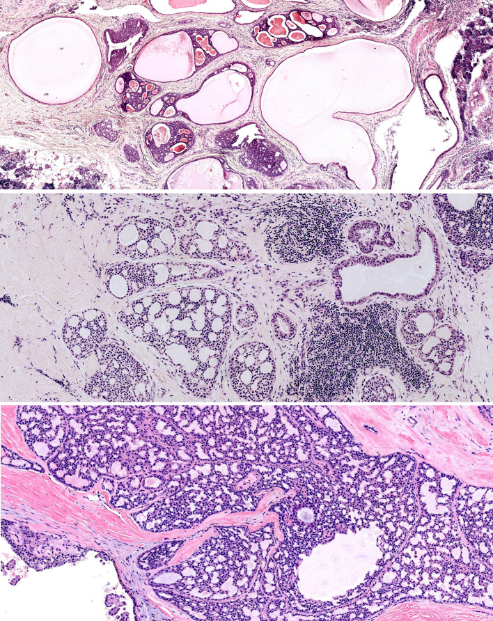
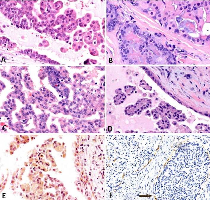
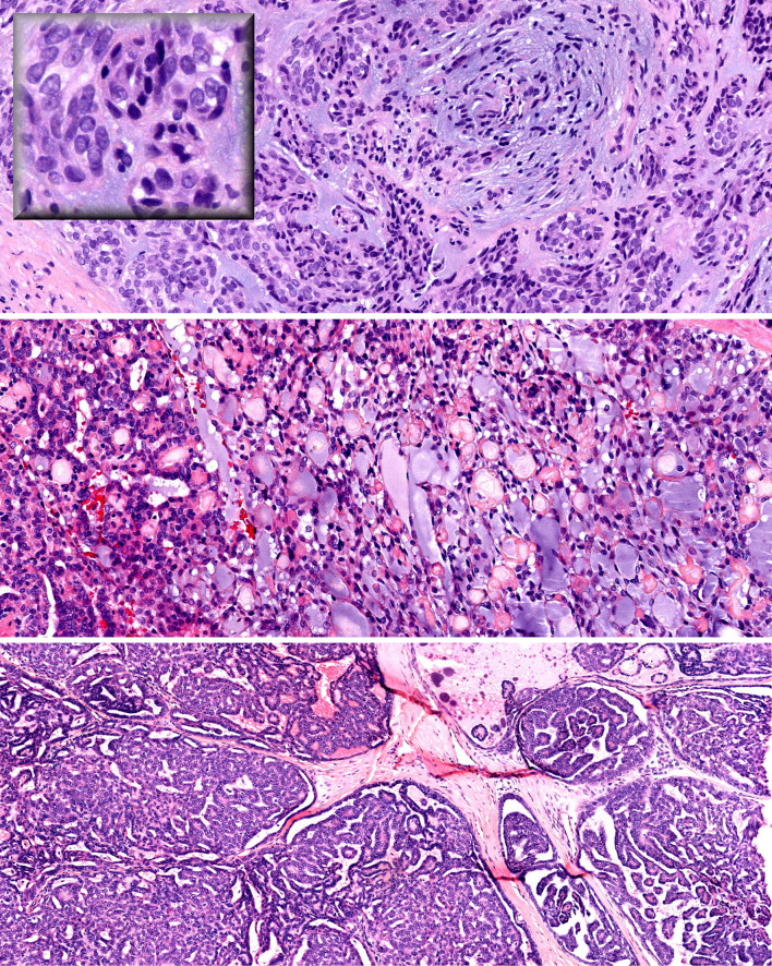
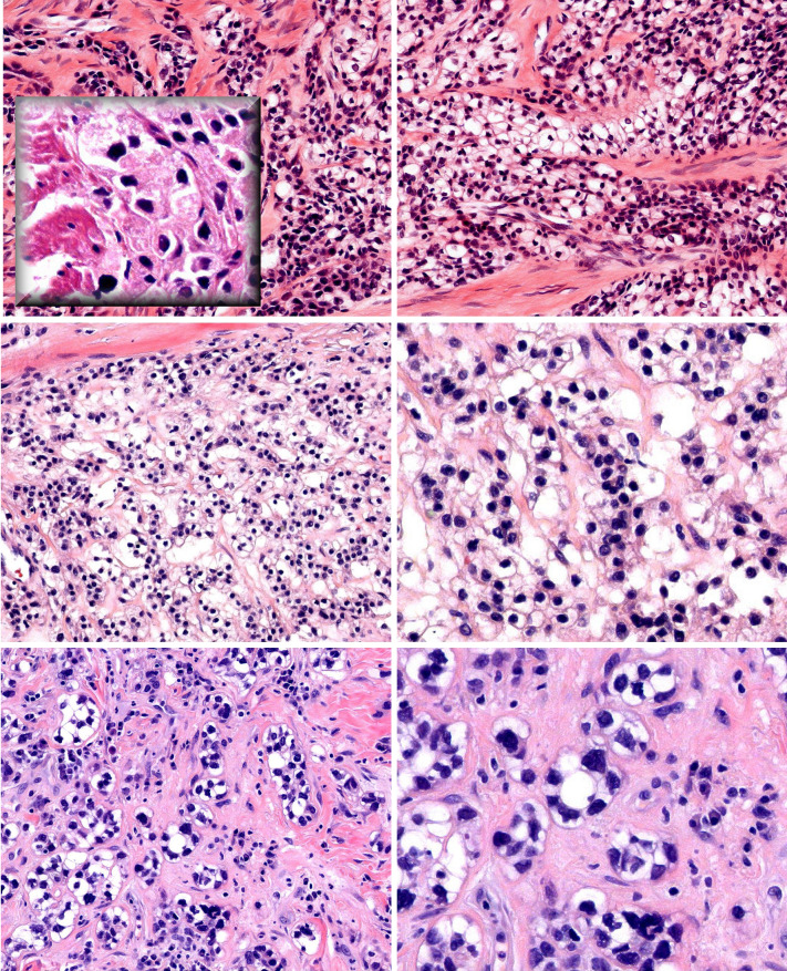
The next WHO classification should abandon "salivary duct carcinoma"; conventional salivary duct carcinoma should be classified as "high-grade salivary duct carcinoma". Low-grade salivary duct carcinoma should replace the current nosology of "low-grade cribriform cystadenocarcinoma". Cystadenocarcinoma should be classified with the descriptor "Not Otherwise Specified" and should be considered an exclusionary diagnostic category. On the other hand, "Not Otherwise Specified" does not fit for hyalinizing clear cell carcinoma (HCCC). The EWSR1-ATF1 fusion is specific for HCCC within the context of salivary neoplasia. We recommend adding "hyalinizing" even though this feature is not present in all cases; the benefit of which is the mental association with a salivary clear cell malignancy. Sinonasal Renal Cell-like Adenocarcinoma (SNRCLA) is a distinct clear cell neoplasm and should be added to the next WHO classification. Future studies will bear out whether SNRCLA is even a low-grade carcinoma, or may be reclassified as "adenoma". Lastly, the next WHO monograph should include the Risk Model in the general introductory statements on oral squamous cell carcinoma, under a subheading of "Histological Prognosticators". The positive predictive value for developing locoregional recurrence in patients with low-stage oral cavity squamous carcinoma (OSCC) and "worst pattern of invasion type-5" (WPOI-5) is 42 %. Low-stage high-risk OSCC with a combination of features other than WPOI-5 is associated with 32 % likelihood for locoregional progression. WPOI-5 also predicts occult metastatic disease (p = 0.0001, Chi squared, 2 DF). Thus the Risk Model can also be used to make decisions regarding staged elective neck dissections.


Figures

Fig. 1
Fig. 1
Low-grade salivary duct carcinoma—cystically dilated ducts with intraductal proliferation (top). Fenestrated, pseudocribriform architecture (middle and bottom)
Fig. 2
Fig. 2
Low-grade salivary duct carcinoma—a through d demonstrate bland tumor cells with papillary or fenestrated architecture. Hobnail cells are seen (a, d). e Demonstrates tumor cells with yellow/brown pigment. f Demonstrates IHC for calponin, which delineates the ducts
Fig. 3
Fig. 3
High-grade salivary duct carcinoma—necrosis (left) and high-grade grade tumor cells (right)
Fig. 4
Fig. 4
Low-grade papillary adenocarcinoma (LGPA) and polymorphous low-grade adenocarcinoma (PLGA) share characteristic features: low-grade epithelioid tumor cells with vesicular nuclei (top inset), whorling pattern and slate blue matrix (top), and lacey pseudocribriform pattern (middle). The bottom panel demonstrates a papillary pattern
Fig. 5
Fig. 5
Hyalinizing clear cell carcinoma—infiltrating cords and nests of clear tumor cells with hyaline production (top). Periodic Acid Schiff reveals granular cytoplasmic glycogen (top inset). A subtle biphasic tumor population is seen composed of cells with less cytoplasm (middle left and right). Nuclear pleomorphism and rare gland formation (bottom left and right)
Fig. 6
Fig. 6
Clear cell odontogenic carcinoma—lobulated growth pattern (top). Strands and nests of clear tumor cells in a myxoid stroma (middle left and right). Cells at the periphery of this nest have less cytoplasm, reminiscent of the second population of smaller tumor cells in HCCC (bottom)
Fig. 7
Fig. 7
Sinonasal renal cell-like adenocarcinoma—gland-forming tumor with pink secretions and increased vascularity reminiscent of CC-RCC (a). Bland tumor cells forming “thyroid-like” follicles (b). Intranuclear holes (c). This SNRCLA was unusual as the bland clear tumor cells were interspersed with cells with basophilic and eosinophilic cytoplasm (d, e, f)

References

    1. Delgado R, Klimstra D, Albores-Saavedra J. Low grade salivary duct carcinoma. A distinctive variant with a low-grade histology and a predominant intraductal growth pattern. Cancer. 1996;78:958–967. doi: 10.1002/(SICI)1097-0142(19960901)78:5<958::AID-CNCR4>3.0.CO;2-8. - DOI - PubMed
    1. Brandwein-Gensler M, Hille J, Wang BY, Urken M, Gordon R, Wang LJ, Simpson JR, Simpson RH, Gnepp DR. Low-grade salivary duct carcinoma: description of 16 cases. Am J Surg Pathol. 2004;28:1040–1044. doi: 10.1097/01.pas.0000128662.66321.be. - DOI - PubMed
    1. Brandwein-Gensler M, Gnepp DR, et al. Low-grade cribriform cystadenocarcinoma. In: Barnes L, Eveson JW, Reichart P, et al., editors. Pathology and genetics head and neck tumors. Lyon: IARC Press; 2005. p. 233.
    1. Kuo YJ, Weinreb I, Perez-Ordonez B. Low-grade salivary duct carcinoma or low-grade intraductal carcinoma? Review of the literature. Head Neck Pathol. 2013;7(Suppl 1):S59–S67. doi: 10.1007/s12105-013-0460-1. - DOI - PMC - PubMed
    1. Wang L, Liu Y, Lin X, Zhang D, Li Q, Qiu X, Wang EH. Low-grade cribriform cystadenocarcinoma of salivary glands: report of two cases and review of the literature. Diagn Pathol. 2013;8:28. doi: 10.1186/1746-1596-8-28. - DOI - PMC - PubMed

MeSH terms