

Current views and perspectives on classification of squamous intraepithelial lesions of the head and neck
- PMID: 24595419
- PMCID: PMC3950392
- DOI: 10.1007/s12105-014-0530-z
Current views and perspectives on classification of squamous intraepithelial lesions of the head and neck
Abstract
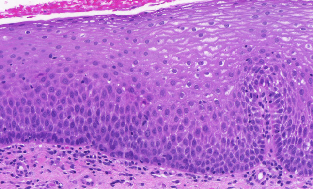
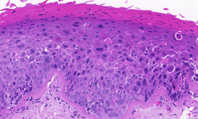
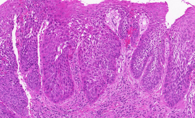
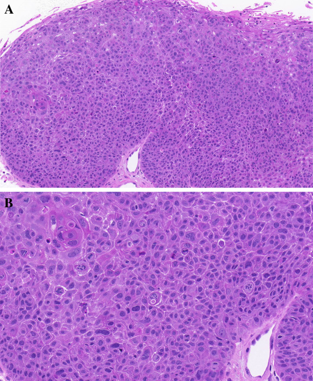
The current state in the field of classifying oral and laryngeal precursor lesions, as proposed in the WHO 2005 Blue Book is not ideal. The results of various inter-observer studies have shown that the currently used grading systems, with different basic concepts and different terminology, cannot continue to be reliably used in the future. The different etiology of cervical and head and neck precursor lesions requires a classification designed to cater to the specificities of the head and neck region. Trying to harmonize different classifications of the oral and laryngeal precursor lesions, we have proposed four crucial steps to set up a unified classification of squamous intraepithelial lesions (SILs): (a) the classification should contain two grades, low-grade and high-grade lesions and, specifically for the larynx, an additional grade-carcinoma in situ (CIS) which must be separated from high-grade laryngeal SILs; (b) the terminology should be unified; our preference is for the term SIL over squamous intraepithelial neoplasia; (c) all leading morphological criteria for low- and high-grade lesions, as well as for CIS, should be clearly defined; (d) agreement between clinicians and pathologists should be achieved on the most appropriate choice of treatment of different grades of SILs in separate head and neck areas.
Figures

References
-
- Gale N, Pilch BZ, Sidransky D, et al. Epithelial precursor lesions. In: Barnes L, Eveson JW, Reichart P, Sidransky D, et al., editors. World Health Organization classification of tumours—pathology and genetics of head and neck tumours. Lyon: IARC Press; 2005. pp. 143–153.
-
- Boy S. Leukoplakia and erythroplakia of oral mucosa—a brief overview. SADJ. 2012;67(10):558–560. - PubMed
-
- Barnes L. Diseases of the larynx, hypopharynx, and trachea. In: Barnes L, editor. Surgical pathology of the head and neck. 3. New York: Informa Healthcare; 2009. pp. 109–200.
MeSH terms
LinkOut - more resources
Full Text Sources
Other Literature Sources
Medical
