

Neuroendocrine neoplasms of the head and neck: some suggestions for the new WHO classification of head and neck tumors
- PMID: 24595420
- PMCID: PMC3950384
- DOI: 10.1007/s12105-014-0531-y
Neuroendocrine neoplasms of the head and neck: some suggestions for the new WHO classification of head and neck tumors
Abstract
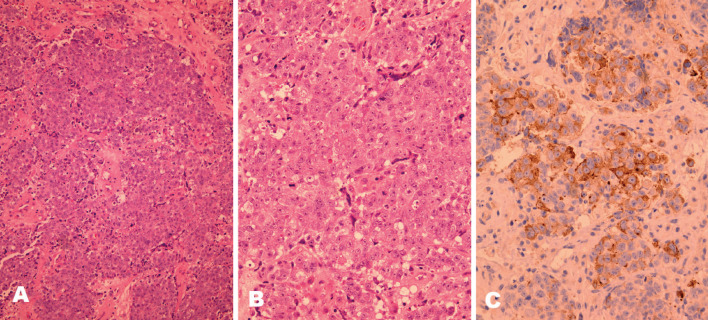
As knowledge and understanding in pathology evolve, classifications and nomenclature also change to reflect those advances. The 2005 World Health Organization Classification of Head and Neck Tumours was a significant step towards diagnostic standardization of head and neck neuroendocrine carcinomas; however, in the last 10 years there have been new data supporting the recognition of "large cell neuroendocrine carcinoma" as a distinctive high grade carcinoma in the head and neck, a lesion not included in the 2005 Classification. In addition, the terms "middle ear adenoma" and "carcinoid tumor of middle ear" are still widely used to describe a neoplasm that is neither a pure adenoma nor a carcinoid tumor but a lesion with variable mixed exocrine and endocrine differentiation. Largely using the diagnostic criteria of the WHO classification of neuroendocrine carcinomas of the lung, we propose the terms "neuroendocrine carcinoma, grade 1"; "neuroendocrine carcinoma, grade 2"; "neuroendocrine carcinoma, grade 3, large cell type"; and "neuroendocrine carcinoma, grade 3, small cell type" for the classification of neuroendocrine carcinomas of the head and neck in a future WHO classification. In addition, we also proposed the term "mixed epithelial neuroendocrine tumor" of the middle ear as an alternative for "middle ear adenoma" and "carcinoid tumor of the middle ear".
Figures


References
-
- Rindi G, Arnold R, Bosman FT, et al. et al. Nomenclature and classification of neuroendocrine neoplams of the digestive system. In: Bosman FT, Carneiro F, Hruban RH, et al.et al., editors. WHO classification of tumours of the digestive system. Lyon: International Agency for Research on Cancer; 2010. pp. 13–14.
-
- Wenig BM, Gnepp DR. The spectrum of neuroendocrine carcinomas of the larynx. Semin Diagn Pathol. 1989;6:329–350. - PubMed
-
- Barnes L, et al. Tumours of the hypopharynx, larynx, and trachea: neuroendocrine tumors. In: Barnes L, Eveson JW, Reichart P, et al., editors. Pathology and genetics head and neck tumours. Lyon: IARC Press; 2005. pp. 135–139.
MeSH terms
LinkOut - more resources
Full Text Sources
Other Literature Sources
Medical
Research Materials
