. 2014 May;62(5):369-88.
doi: 10.1369/0022155414528514. Epub 2014 Mar 4.

Brain barriers and a subpopulation of astroglial progenitors of developing human forebrain are immunostained for the glycoprotein YKL-40

Affiliations

Brain barriers and a subpopulation of astroglial progenitors of developing human forebrain are immunostained for the glycoprotein YKL-40

Camilla Bjørnbak et al. J Histochem Cytochem. 2014 May.

Abstract

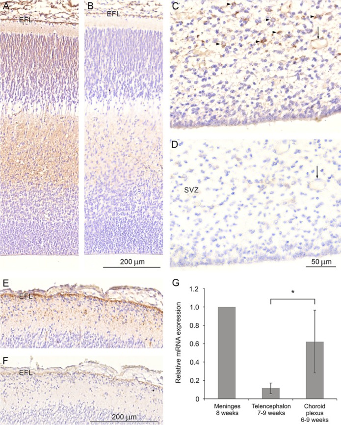
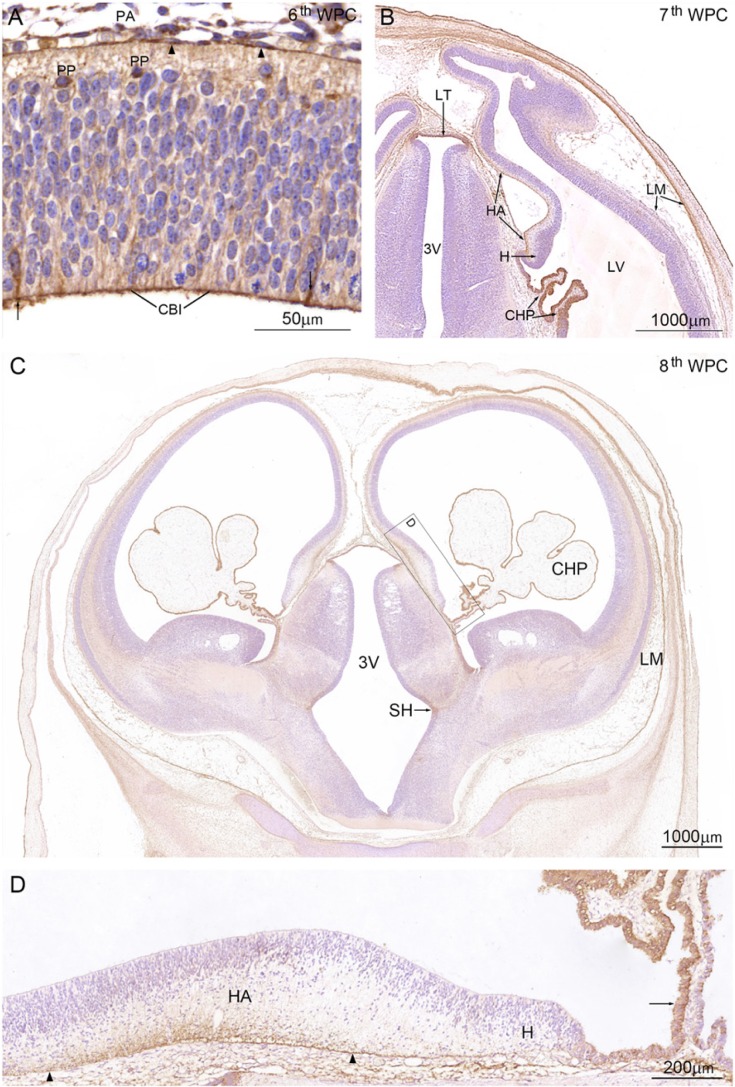
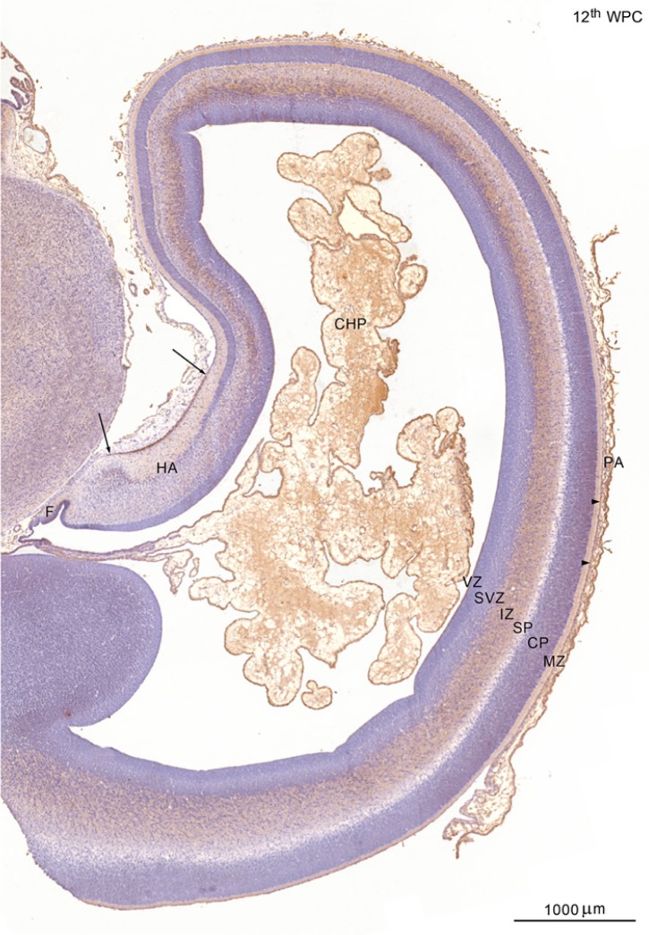
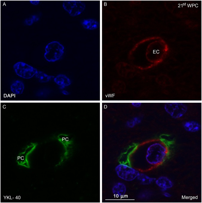
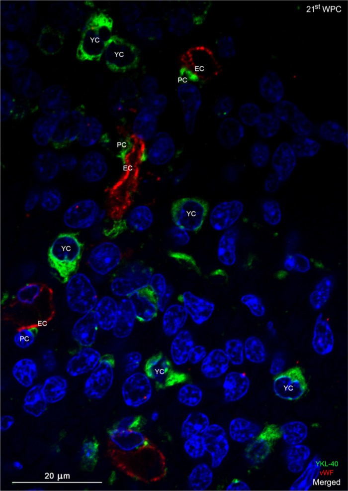
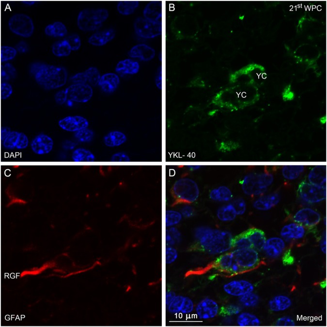
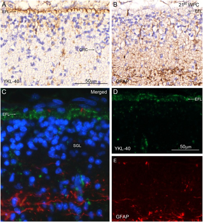
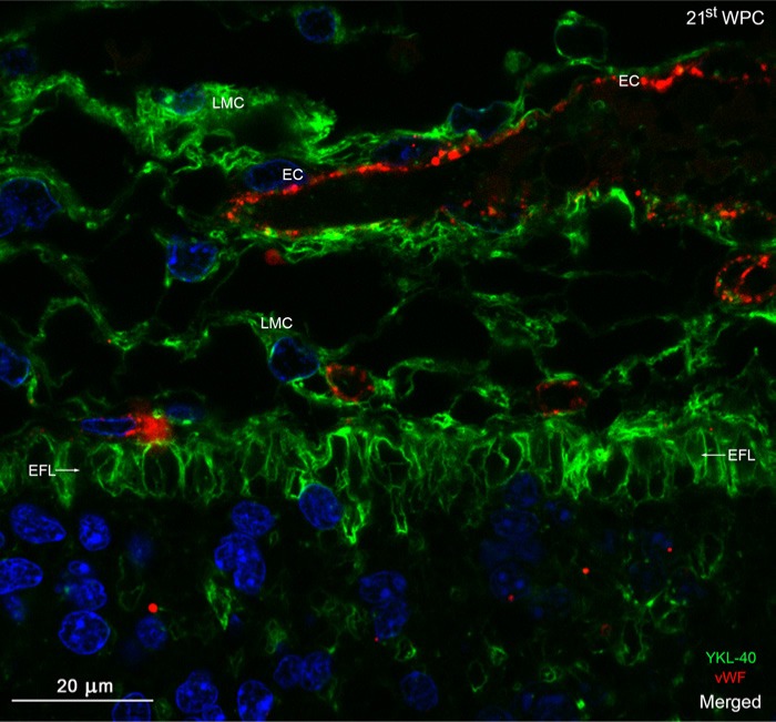
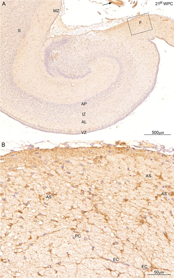
YKL-40, a glycoprotein involved in cell differentiation, has been associated with neurodevelopmental disorders, angiogenesis, neuroinflammation and glioblastomas. We evaluated YKL-40 protein distribution in the early human forebrain using double-labeling immunofluorescence and immunohistochemistry. Immunoreactivity was detected in neuroepithelial cells, radial glial end feet, leptomeningeal cells and choroid plexus epithelial cells. The subpial marginal zone was YKL-40-positive, particularly in the hippocampus, from an early beginning stage in its development. Blood vessels in the intermediate and subventricular zones showed specific YKL-40 reactivity confined to pericytes. Furthermore, a population of YKL-40-positive, small, rounded cells was identified in the ventricular and subventricular zones. Real-time quantitative RT-PCR analysis showed strong YKL-40 mRNA expression in the leptomeninges and the choroid plexuses, and weaker expression in the telencephalic wall. Immunohistochemistry revealed a differential distribution of YKL-40 across the zones of the developing telencephalic wall. We show that YKL-40 is associated with sites of the brain barrier systems and propose that it is involved in controlling local angiogenesis and access of peripheral cells to the forebrain via secretion from leptomeningeal cells, choroid plexus epithelium and pericytes. Furthermore, we suggest that the small, rounded, YKL-40-positive cells represent a subpopulation of astroglial progenitors, and that YKL-40 could be involved in the differentiation of a particular astrocytic lineage.

Keywords: astrocytes; brain barriers; cerebral cortex; development; neural stem cells.


Conflict of interest statement

Declaration of Conflicting Interests: The author(s) declared no potential conflicts of interest with respect to the research, authorship, and/or publication of this article.

Figures

Figure 1.
Figure 1.
YKL-40 protein distribution in the neocortex of a human fetus at the 12th week post-conception (wpc) (CRL: 75 mm) (A-B), and in the occipital cortex of a 21st wpc fetus (CRL: 200 mm) (C-F). Sections shown in (A), (C) and (E) were stained with the monoclonal antibody (MAb) 201.F9 whereas those in (B), (D) and (F) were stained using the MAb 201.F9 preabsorbed with purified human YKL-40 protein. Note the lack of or a markedly diminished reactivity of the sections stained with the preabsorbed YKL-40 antibody. The end feet layer (EFL) is barely visible in (B) and (F), and the presumed astroglial progenitors (arrow) in the subventricular zone (SVZ) in (C) is no longer positive following preabsorption (D). Note the reactivity associated with the blood vessel in (C) (arrow) and the lack of reactivity in (D) (arrow). (G) Quantitative real-time RT-PCR analysis of YKL-40 mRNA expression in the human developing forebrain from tissue samples dissected from two human embryos (aged 6 weeks and 5 days post-conception and 7 wpc) at the 7th and 8th wpc and three human fetuses (aged 8 wpc, 8 weeks and 3 days post-conception, and 9 wpc) at the 9th and 10th wpc. Relative mRNA expression is shown (meninges=1). Only one meninges sample was available for analysis. A significant difference between average expression values obtained in the telencephalon (n=4) and the choroid plexus (n=3) was observed (indicated by *, Students t-test, p=0.029). Error bars represent one standard deviation. Scale bar: A–B: 200 μm, C–D: 50 μm, E–F: 200 μm. Same magnification in (A-B), (C-D) and (E-F).
Figure 2.
Figure 2.
YKL-40 protein distribution in a coronal section from an embryo at the 6th wpc (crown-rump length (CRL): 7 mm). (A) Leptomeningeal cells (arrowheads) in the pia-arachnoid (PA) have strong YKL-40 immunoreactivity. Apical surfaces of neuroepithelial cells forming the CSF-brain interface (CBI) are outlined by YKL-40 immunoreactivity and some neuroepithelial cells/radial glial cells (arrows) and preplate cells (PP) are YKL-40 positive. (B) Coronal section from a 7th wpc human embryo (CRL: 20 mm). The YKL-40 positive outer surface of the hippocampal anlage (HA) is shown between the two arrows. Epithelial cells of the newly-formed lateral ventricular choroid plexus (CHP) have strong YKL-40 immunoreactivity. The cortico-choroid boundary between the hem (H) and the plexus is YKL-40 positive. Leptomeninges (LM), in particular, the continuous part of the arachnoid, show strong YKL-40 immunoreactivity. In the rostral part of third ventricle (3V) the lamina terminalis (LT) shows strong YKL-40 immunoreactivity. (C) Coronal section from a late 8th wpc human embryo (CRL: 31 mm). Note the distinct reactivity of the LM and CHP epithelium compared with that of the telencephalic wall, where only marginal and intermediate zones show a weak YKL-40 immunoreactivity. A prominent sulcus hypothalamicus (SH) is seen in 3V. The boxed area includes the hippocampal anlage (HA), the hem (H) and the root of the choroid plexus, and is shown in higher magnification in (D). End feet and the outer marginal zone of the developing CA1 to CA4 of the hippocampus (between the arrowheads) show strong YKL-40 immunoreactivity. The dentate primordium between the right arrowhead and the hem is only immunostained in the end feet region in contrast to the unstained hem, which extends to the choroid plexus where the part of the root (arrow) adjacent to the hem shows strong YKL-40 immunoreactivity. Scale bars: A: 50 μm; B-C: 1000 μm; D: 200 μm.
Figure 3.
Figure 3.
YKL-40 protein distribution in the neocortex and hippocampus of a 12th wpc fetus (crown-rump length: 75 mm). The pia-arachnoid (PA) immediately outside the telencephalic wall and of the choroid plexus (CHP) on the inside show strong YKL-40 immunoreactivity. Gradients within the wall include a marked staining of the outermost layer of the marginal zone (MZ) of the hippocampal anlage (HA) between the two arrows, and a consistent reactivity of the subpial line of the lateral neocortex (arrowheads). The cortical plate (CP) and subplate (SP) are not stained in contrast with the intermediate zone (IZ), which is strongly reacting from the subiculum via the medial (cingulate) cortex to the dorsal neocortex. The subventricular zone (SVZ) and ventricular zone (VZ) have no YKL-40 immunoreactivity at this low magnification except from the positively stained VZ covering the fimbria (F). Scale bar: 1000 μm.
Figure 4.
Figure 4.
YKL-40 protein distribution in the lateral parietal forebrain of a 15th wpc human fetus (crown-rump length: 111 mm). (A) The inner half of the cerebral wall has small dots of YKL-40 immunoreactivity in the intermediate zone (IZ), in contrast to the subventricular and ventricular zones (SVZ and VZ) which show virtually no YKL-40 immunoreactivity at this low magnification. (B) A higher magnification of the VZ–SVZ, with virtually no YKL-40 immunoreactivity. A small vessel with a YKL-40 positive endothelial cell/pericyte is seen in the lower part of the IZ (arrows). (C) Higher magnification of the IZ boxed area in (A). The fine granular staining of YKL-40 is associated with small blood vessels (arrows), but whether the reaction product is within endothelial cells and/or pericytes cannot be distinguished. A longitudinal vessel with patchy reactivity is seen (arrowheads). Astrocytes with end feet are not developed at this stage. Scale bars: A: 200 μm; B-C: 50 μm.
Figure 5.
Figure 5.
Confocal image of a double immunolabeled section of the occipital intermediate zone from a 21st wpc human fetus (crown-rump length: 200 mm) stained with antibodies against YKL-40 (green) and von Willebrand Factor (vWF; red) and nuclear counterstained with DAPI (blue). (A-D) Microvessel with an endothelial cell (EC) and surrounding pericytes (PC). (B) vWF-positive endothelial lining of an intermediate zone capillary and the EC. (C) Discontinuity of the surrounding YKL-40-positive PC. (D) Merged image showing the lack of co-localization of vWF and YKL-40 in the YKL-40-positive PC membrane adjacent to the endothelial lining. Scale bar: 10 μm.
Figure 6.
Figure 6.
Confocal image of a double immunolabeled section of the occipital subventricular zone from a 21st wpc human fetus (crown-rump length: 200 mm) stained with antibodies against YKL-40 (green) and von Willebrand Factor (vWF; red) and nuclear counterstained with DAPI (blue). The YKL-40-immunopositive pericytes (PC) in the subventricular zone show a circumventing discontinuity of the vWF-immunopositive endothelial cells (EC) of the microvessels. YKL-40 immunoreactivity is found in small rounded cells (YKL-40-positice cells, YC) with a strong cytoplasmic staining surrounding the nucleus. These cells have no contact with microvessels. Scale bar: 20 μm.
Figure 7.
Figure 7.
Distribution of YKL-40 and GFAP in the occipital subventricular zone from a 21st wpc human fetus (crown-rump length: 200 mm). Sections were stained with antibodies against YKL-40 (green) and glial fibrillary acidic protein (GFAP, red), counterstained with DAPI (blue) and examined in the confocal microscope throughout the z-axis. (A) DAPI. (B) YKL-40-immunopositive small rounded cells (YC). (C) GFAP-immunopositive radial glial fibers (RGF). In (D), note the co-localization between the GFAP-positive RGF and YKL-40. Several GFAP-positive YCs are seen with a strong cytoplasmic YKL-40 reactivity. Scale bar: 10 μm.
Figure 8.
Figure 8.
Distribution of YKL-40 and GFAP in the marginal zone from the dorso-lateral parietal cortex of a 21st wpc human fetus (crown-rump length: 200 mm). Consecutive sections are stained with antibodies against YKL-40 and glial fibrillary acidic protein (GFAP), nuclear counterstained with hematoxylin (A, B) or DAPI (C) and viewed with ordinary light microscopy (A-B) or confocal microscopy (C-E). In (A) and (D) the end feet layer (EFL) shows strong YKL-40 immunoreactivity; however, virtually no GFAP staining is seen in the EFL in (B) and (E). The large Cajal-Retzius cell (CRC) in (A) has no YKL-40 immunoreactivity. The marginal zone contains a transient population of YKL-40-negative cells corresponding to the subpial granular layer (SGL) shown with DAPI staining in the merged image in (C). Scale bars: A-E: 50 μm; the same magnification was used in (A-B), and in (C-E).
Figure 9.
Figure 9.
Distribution of YKL-40 and von Willebrand factor in the occipital marginal zone from a 21st wpc human fetus (crown-rump length: 200 mm). The section was stained with antibodies against YKL-40 (green) and von Willebrand Factor (vWF) (red), nuclear counterstained with DAPI (blue) and viewed using confocal microscopy. The YKL-40-immunopositive end feet layer (EFL, arrows) separates the leptomeninges and underlying marginal zone. No blood vessels penetrate the marginal zone in this section. Note the strongly immunoreactive leptomeningeal cells (LMC), some of which surround the vWF-positive endothelial cells (EC) of the vessels of the pia-arachnoid. The EFL forming the outermost part of the marginal zone shows a strong membranous reactivity, with a decreasing immunoreactivity within its deeper strata. Scale bar:20 μm.
Figure 10.
Figure 10.
Distribution of YKL-40 in a coronal section of the hippocampus from a 21st wpc human fetus (crown-rump length: 200 mm). (A) In contrast to the weak staining of the hippocampus, the fimbria (F)—shown at higher magnification in (B)—and smooth muscle cells (arrow) in blood vessels in the subarachnoid space show strong YKL-40 immunoreactivity. The subpial layer of the marginal zone (MZ) of the subiculum (S) and the cellular layer between alveus (AL) and the ventricular zone (VZ) are also reactive. The intermediate zone (IZ) facing the ammonic plate (AP) shows very small dots of YKL-40 immunoreactivity. In (B), which is a larger magnification of the boxed area in (A), many YKL-40-immunopositive astrocyte-resembling cells (AS)form a network with endothelial cells (EC) and pericytes (PC). Scale bars: A: 500 μm; B: 50 μm.

References

    1. Abraham H, Pérez-García CG, Meyer G. (2004). P73 and Reelin in Cajal-Retzius Cells of the Developing Human Hippocampal Formation. Cereb Cortex 14:484-495 - PubMed
    1. Aguzzi A, Barres BA, Bennett ML. (2013). Microglia: scapegoat, saboteur, or something else? Science 339:156-161 - PMC - PubMed
    1. Alvarez-Buylla A, Seri B, Doetsch F. (2002). Identification of neural stem cells in the adult vertebrate brain. Brain Res Bull 57:751-758 - PubMed
    1. Argaw AT, Asp L, Zhang J, Navrazhina K, Pham T, Mariani JN, Mahase S, Dutta DJ, Seto J, Kramer EG, Ferrara N, Sofroniew MV, John GR. (2012). Astrocyte-derived VEGF-A drives blood-brain barrier disruption in CNS inflammatory disease. J Clin Invest 122:2454-2468 - PMC - PubMed
    1. Arion D, Unger T, Lewis DA, Levitt P, Mirnics K. (2007). Molecular evidence for increased expression of genes related to immune and chaperone function in the prefrontal cortex in schizophrenia. Biol Psychiatry 62:711-721 - PMC - PubMed

Publication types

LinkOut - more resources