. 2014 Apr;144(3):539-49.
doi: 10.1007/s10549-014-2896-8. Epub 2014 Mar 5.

Immune cell quantitation in normal breast tissue lobules with and without lobulitis

Affiliations

Immune cell quantitation in normal breast tissue lobules with and without lobulitis

Amy C Degnim et al. Breast Cancer Res Treat. 2014 Apr.

Abstract

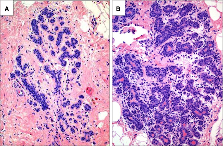
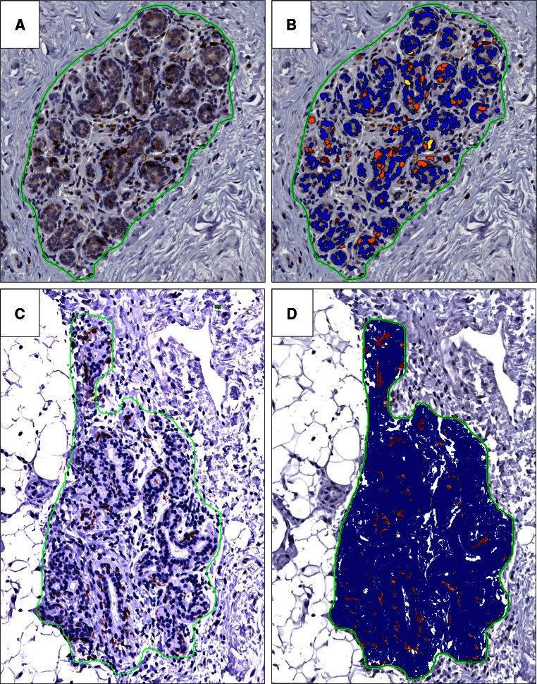
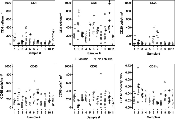
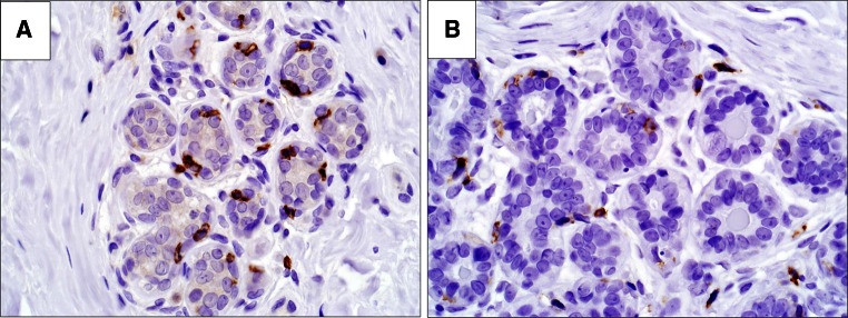
While the immune microenvironment has been investigated in breast cancers, little is known about its role in non-malignant breast tissues. Here we quantify and localize cellular immune components in normal breast tissue lobules, with and without visible immune infiltrates (lobulitis). Up to ten representative lobules each in eleven normal breast tissue samples were assessed for B cells (CD20), cytotoxic T cells (CD8), helper T cells (CD4), dendritic cells (CD11c), leukocytes (CD45), and monocytes/macrophages (CD68). Using digital image analysis, immune cell densities were measured and compared between lobules with/without lobulitis. 109 lobules in 11 normal breast tissue samples were evaluated; 31 with lobulitis and 78 without. Immune cells showed consistent patterns in all normal samples, predominantly localized to lobules rather than stroma. Regardless of lobulitis status, most lobules demonstrated CD8+, CD11c+, CD45+, and CD68+ cells, with lower densities of CD4+ and CD20+ cells. Both CD11c+ and CD8+ cells were consistently and intimately associated with the basal aspect of lobule epithelium. Significantly higher densities of CD4+, CD8+, CD20+, and CD45+ cells were observed in lobules with lobulitis. In contrast, densities of monocytes/macrophages and dendritic cells did not vary with lobulitis. In normal breast tissue, myeloid and lymphoid cells are present and localized to lobules, with cytotoxic T and dendritic cells directly integrated with epithelium. Lobules with lobulitis have significantly more adaptive immune (B and T) cells, but no increase in dendritic cells or monocytes/macrophages. These findings indicate an active and dynamic mucosal immune system in normal breast tissue.


Figures

Fig. 1
Fig. 1
The presence of immune infiltrates that define lobulitis. a Normal lobule, without lobulitis; b normal lobule, with lobulitis. H&E stain, ×200 magnification
Fig. 2
Fig. 2
Quantification of immune cells in normal lobules. a, b CD8+ cell quantitation in a normal lobule without lobulitis. a immunostain alone, b color overlay demonstrating positively stained cells within circled lobule; positive cells are identified by red and orange color, yellow cells are below staining threshold for positivity, and blue cells are non-staining cells. c, d CD11c quantitation by pixel count ratio in a normal lobule without lobulitis, ×200 magnification. c Immunostain alone, d color overlay demonstrating pixels with positive staining (red and orange) compared to non-stained pixels (blue). ×200 magnification
Fig. 3
Fig. 3
Quantitative immune cell densities for individual lobules in each of the 11 subjects studied; open circles indicate lobules without lobulitis, and crosses indicate lobules with lobulitis. Asterisk in CD8 density plot denotes one point outside the plotting region for subject #10 (value was 1336 CD8 cells/mm2)
Fig. 4
Fig. 4
Direct association of dendritic cells and CD8 T cells within epithelium. a CD8 positive cells located at the basal aspect of the epithelium, b CD11c positive staining extending more diffusely around epithelium. ×400 magnification

References

    1. Ghajar CM. On leukocytes in mammary development and cancer. Cold Spring Harbor Perspect Biol. 2012 - PMC - PubMed
    1. Ghajar CM, Bissell MJ. Extracellular matrix control of mammary gland morphogenesis and tumorigenesis: insights from imaging. Histochem Cell Biol. 2008;130(6):1105–1118. doi: 10.1007/s00418-008-0537-1. - DOI - PMC - PubMed
    1. Reed JR, Schwertfeger KL. Immune cell location and function during post-natal mammary gland development. J Mammary Gland Biol Neoplasia. 2010;15(3):329–339. doi: 10.1007/s10911-010-9188-7. - DOI - PMC - PubMed
    1. Brandtzaeg P. The mucosal immune system and its integration with the mammary glands. J Pediatr. 2010;156(2 Suppl):S8–S15. doi: 10.1016/j.jpeds.2009.11.014. - DOI - PubMed
    1. Goldman AS. The immune system of human milk: antimicrobial, antiinflammatory and immunomodulating properties. Pediatr Infect Dis J. 1993;12(8):664–671. doi: 10.1097/00006454-199308000-00008. - DOI - PubMed

Publication types

MeSH terms