

Comment
doi: 10.7554/eLife.02369.
Three-stranded antiviral attack
Affiliations
- PMID: 24596154
- PMCID: PMC3941025
- DOI: 10.7554/eLife.02369
Item in Clipboard
Comment
Three-stranded antiviral attack
Elife.
.
Abstract
Mitochondrial antiviral signalling proteins form an intricate three-stranded helical filament that has a central role in the response of cells to viruses.
Keywords: MAVS; cryoEM reconstruction; immune response; innate immunity; prion-like filaments; three-stranded filaments.
Conflict of interest statement
Figures
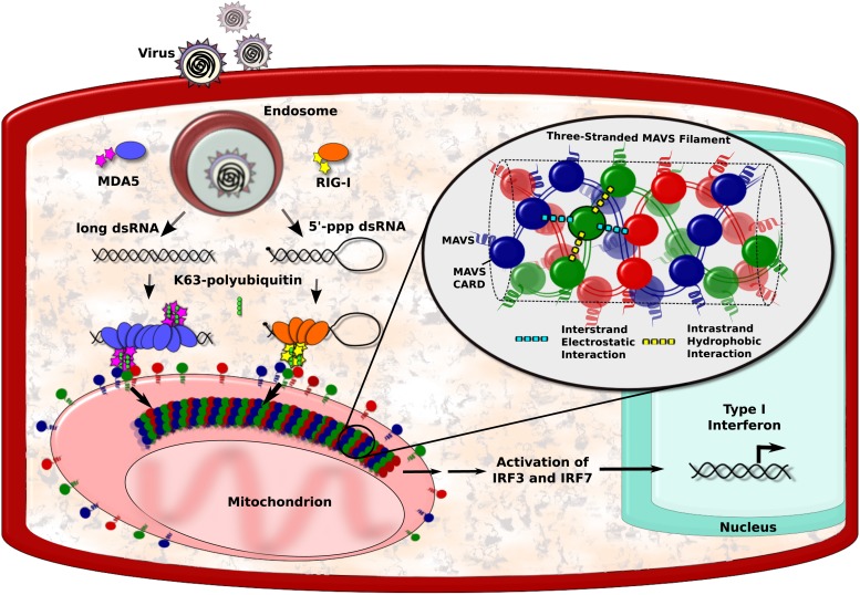
Viral infection introduces foreign RNA molecules into cells (top left), where they are detected by various sensors. A sensor called RIG-I binds to RNA molecules that contain an exposed 5′-triphosphate and terminal dsRNA structure, while MDA5 binds to long dsRNA molecules (Goubau and Deddouche, 2013). The complexes formed by the RNA molecules and the sensors then activate MAVS (mitochondrial antiviral signalling) proteins that are found on the surface of mitochondria. This occurs in the presence of polyubiquitin, which serves to bridge and stabilise the complexes (Jiang et al., 2012). Binding between the complexes and the MAVS proteins occurs at regions on each named CARDs (caspase activation and recruitment domains). In the figure, the RIG-I and MDA5 CARDs are shown as stars, and the MAVS CARDs by the red, blue and green circles. This binding organises the MAVS proteins into filaments to form a signalling platform that activates transcription factors (IRF3 and IRF7), which go on to stimulate the transcription of antiviral type I interferon (IFN-I) genes. Xu et al. report that each MAVS filament contains three intertwined helical strands (shown in red, blue and green). The structure is maintained by interactions between the MAVS CARDs: each CARD in a filament interacts with two other CARDs in the same strand and two CARDs in neighbouring strands. Interactions between strands are hydrophobic (cyan dashed lines), and interactions within a strand are electrostatic (yellow dashed lines).
Comment on
-
Structural basis for the prion-like MAVS filaments in antiviral innate immunity.Elife. 2014 Jan 1;3:e01489. doi: 10.7554/eLife.01489. Elife. 2014. PMID: 24569476 Free PMC article.
References
Publication types
MeSH terms
Substances
LinkOut - more resources
Full Text Sources
Other Literature Sources
Miscellaneous
