Review
. 2014 Apr 16;33(8):788-822.
doi: 10.1002/embj.201386357. Epub 2014 Mar 3.

Synaptic vesicle recycling: steps and principles

Affiliations
Review

Synaptic vesicle recycling: steps and principles

Silvio O Rizzoli. EMBO J. .

Abstract

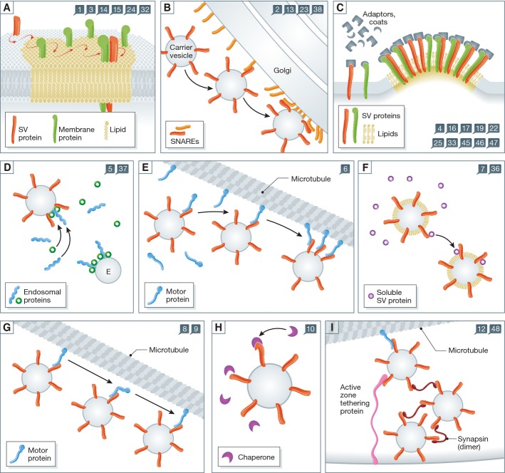
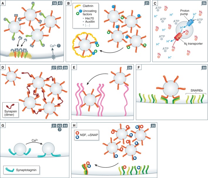
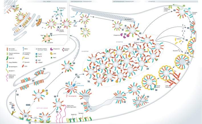
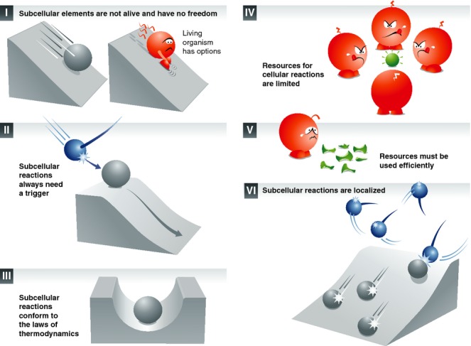
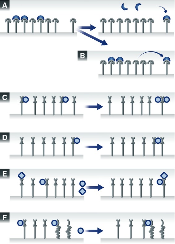
Synaptic vesicle recycling is one of the best-studied cellular pathways. Many of the proteins involved are known, and their interactions are becoming increasingly clear. However, as for many other pathways, it is still difficult to understand synaptic vesicle recycling as a whole. While it is generally possible to point out how synaptic reactions take place, it is not always easy to understand what triggers or controls them. Also, it is often difficult to understand how the availability of the reaction partners is controlled: how the reaction partners manage to find each other in the right place, at the right time. I present here an overview of synaptic vesicle recycling, discussing the mechanisms that trigger different reactions, and those that ensure the availability of reaction partners. A central argument is that synaptic vesicles bind soluble cofactor proteins, with low affinity, and thus control their availability in the synapse, forming a buffer for cofactor proteins. The availability of cofactor proteins, in turn, regulates the different synaptic reactions. Similar mechanisms, in which one of the reaction partners buffers another, may apply to many other processes, from the biogenesis to the degradation of the synaptic vesicle.


Figures

Figure 1
Figure 1. Different types of reactions that occur during vesicle recycling
The numbers in the blue squares indicate the reaction steps addressed by the schemes. (A) A SV protein has entered an organelle and is sorting in the membrane. It encounters a domain of lipids and proteins that it has a limited affinity for, and diffuses slowly within the domain. It eventually becomes stabilized within the domain by binding multiple partners (lipids, proteins). (B) A carrier vesicle fuses with an organelle, such as the Golgi apparatus. The vesicle is buffered locally by the surface of the Golgi apparatus, which is enriched in molecules involved in docking and tethering carrier vesicles, as well as in the fusion with these vesicles (SNARE proteins). The meeting of fusion molecules from the two membranes triggers the fusion of the organelles. (C) Budding reactions. The accumulation of several types of SV molecules in a membrane domain triggers the recruitment of several types of adaptor and coat proteins, each binding to its own preferred SV partner. The accumulation of the coats and adaptors eventually surpasses a critical mass and thus induces the budding reaction. (D) Processing through an endosome. Components of the endosomal pathway, such as Rabs and their effectors, are recruited to carrier vesicles, by interacting with multiple components (proteins, lipids) of the vesicles. This gives the carrier vesicles and endosomal nature, and allows them to fuse to endosomes. Note that this is a putative step in SV recycling. (E) The carrier vesicle interacts with a motor protein, whose high affinity for microtubules causes the eventual delivery of the carrier to the microtubule bundle. Here it may bind further motors, and may proceed along the microtubule toward the synapse. (F) Soluble proteins are recruited onto the carrier vesicle, by interacting with its protein and lipid components. (G) Progression of the vesicle along microtubules (anterograde transport). (H) Recruitment of chaperone proteins onto the carrier vesicle. The chaperone is buffered by the vesicle through low-affinity interactions with normal proteins, until binding more strongly to a spontaneously unfolded protein. In this way the chaperones “probe” continually the surface of the vesicle, and can rapidly and efficiently detect unfolded elements. (I) The carrier vesicle comes off the microtubule track, by interacting with docking proteins and/or with other “sticky” proteins such as synapsin. These multiple interactions are stronger than the interaction with the motor, and remove the vesicle from the microtubule.
Figure 2
Figure 2. More types of reaction schemes from vesicle recycling
As in Fig 1, the numbers in the blue squares indicate the reaction steps addressed by the schemes. (A) In order for endocytosis to happen, adaptor and coat proteins need to be recruited from a source within the synapse. This source may be the SV cluster: many adaptors and coat proteins may be bound onto SV proteins at rest. They may be released during activity and will participate in endocytosis. (B) Similar to panel A, uncoating factors may be recruited from the vesicle cluster onto a coated vesicle. (C) The reactions involved in neurotransmitter refilling: vATPase molecules acidify the SV, and neurotransmitter molecules enter it. (D) The SV may become entangled in a synapsin meshwork, by spontaneous binding to one or more synapsin molecules. (E) Docking at the active zone – in the same fashion as in panel D, but through interactions with active zone proteins. (F) The SV engages plasma membrane SNAREs and prepares (in a sense) for fusion. The reaction is relatively similar to the one from panels D-E, with SNAREs being the interacting molecules. (G) Calcium stimulates fusion by interactions with sensor proteins such as synaptotagmin. (H) As in panels A or B, α-SNAP and NSF are recruited onto SNARE complexes either from the vesicle cluster or from SNAREs on the plasma membrane. The result is the separation of SNARE complexes.
Figure 3
Figure 3. Overview of synaptic vesicle recycling
New SV proteins are generated in the ER and diffuse to specific domains (step 1), before budding and fusion of the carrier vesicle to the Golgi apparatus (step 2). Sorting in the Golgi apparatus, in which some contaminant molecules are removed (orange; step 3), is followed by budding from the Golgi apparatus (step 4). The new carrier vesicle may sort through an endosome (step 5), and will interact with motor proteins to reach microtubules (step 6). Soluble SV proteins may bind specific components of the carrier and be transported along (step 7). Other proteins may also tag along, such as chaperones (step 10), through non-specific interactions with the carrier vesicle proteins. Anterograde transport follows (step 8); it will be blocked if any damage to microtubules takes place (step 9). It is doubtful whether there is any fusion between carrier vesicles along the way (step 11). The carrier eventually comes off microtubules in the synapse (step 12), and will fuse to the plasma membrane (step 13). Sorting of contaminants follows (green, step 14), in parallel with recruitment of other SV proteins (blue, step 15). Budding from the plasma membrane follows (steps 16, 17, 18 and 19), and the coated vesicle is pushed by actin away from the membrane (step 20), before uncoating (step 21). The newly uncoated vesicles do not fuse to each other (step 22), but may fuse to an endosome (step 23), which is followed by endosomal sorting (step 24) and budding (step 25). The new SV fills with neurotransmitter (step 26). The SV either remains mobile for a while (steps 27, 29), docks at the active zone (step 28), or becomes integrated in the SV cluster (step 39). Priming for fusion (step 30) follows docking, and in turn is followed by fusion, upon action potential stimulation and calcium entry (step 31) or in spontaneous fashion, independent of stimulation (step 43). Sorting of SV components may happen in the plasma membrane, to be eventually followed by endocytosis (steps 32, 33). Before endocytosis the SNARE complexes that formed during fusion are separated (step 35), which is an important sorting step for SV components. Damaged SV proteins may be targeted by the proteasomal system (step 36; note that this reaction is likely to happen almost exclusively for soluble SV proteins, although for simplicity the SV protein is depicted here on the SV membrane). Damaged SVs may be tagged for retrograde transport (step 37). Lysosomal degradation awaits (step 38). The SV cluster forms a reserve for the various soluble proteins involved in SV recycling (step 42). Finally, strong synaptic stimulation results in massive exocytosis (step 44) and formation of membrane infoldings (step 45), from which endocytosis machinery removes SV-sized chunks (step 46).
None
None

References

    1. Abraham C, Hutter H, Palfreyman MT, Spatkowski G, Weimer RM, Windoffer R, Jorgensen EM, Leube RE. Synaptic tetraspan vesicle membrane proteins are conserved but not needed for synaptogenesis and neuronal function in Caenorhabditis elegans. Proc Natl Acad Sci USA. 2006;103:8227–8232. - PMC - PubMed
    1. Ahmari SE, Buchanan J, Smith SJ. Assembly of presynaptic active zones from cytoplasmic transport packets. Nat Neurosci. 2000;3:445–451. - PubMed
    1. Ahnert-Hilger G, Höltje M, Pahner I, Winter S, Brunk I. Regulation of vesicular neurotransmitter transporters. Rev Physiol Biochem Pharmacol. 2003;150:140–160. - PubMed
    1. Alabi AA, Tsien RW. Perspectives on Kiss-and-Run: role in exocytosis, endocytosis, and neurotransmission. Annu Rev Physiol. 2013;75:393–422. - PubMed
    1. Andreae LC, Fredj NB, Burrone J. Independent vesicle pools underlie different modes of release during neuronal development. J Neurosci. 2012;32:1867–1874. - PMC - PubMed

Publication types

Substances