Review
. 2014 Mar;31(1):33-41.
doi: 10.1055/s-0033-1363841.

Percutaneous ablation of the small renal mass-techniques and outcomes

Affiliations
Review

Percutaneous ablation of the small renal mass-techniques and outcomes

Andrew J Gunn et al. Semin Intervent Radiol. 2014 Mar.

Abstract

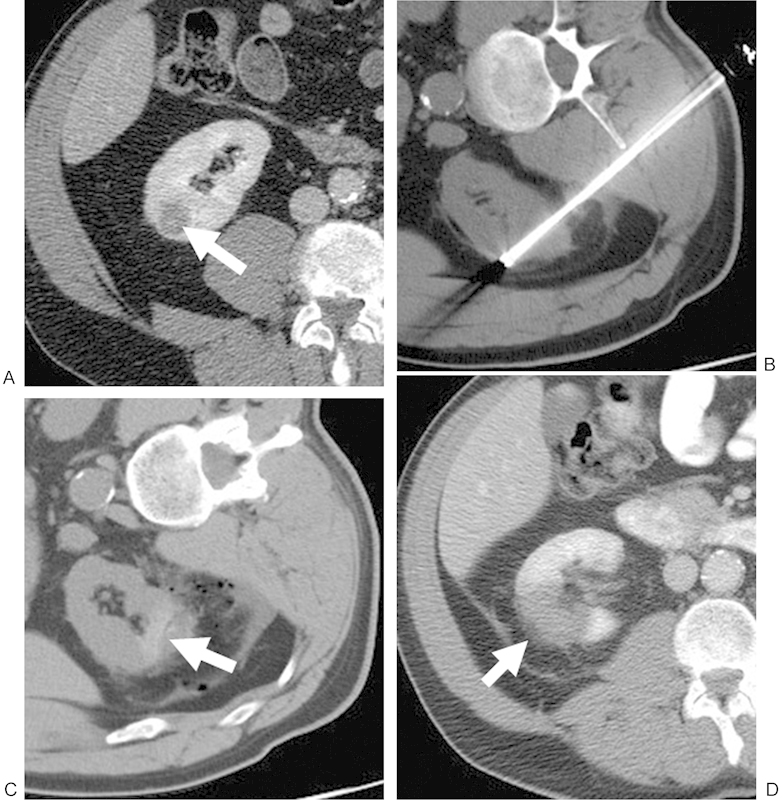
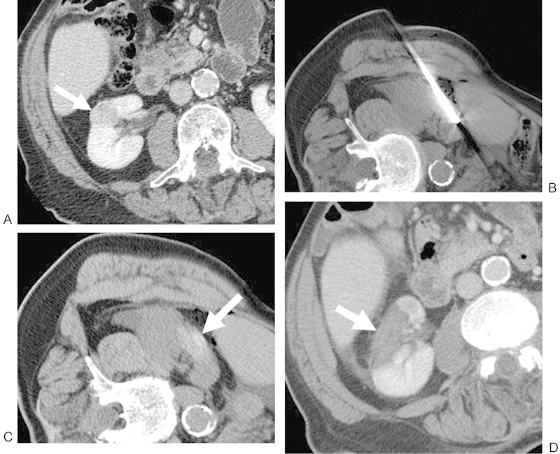
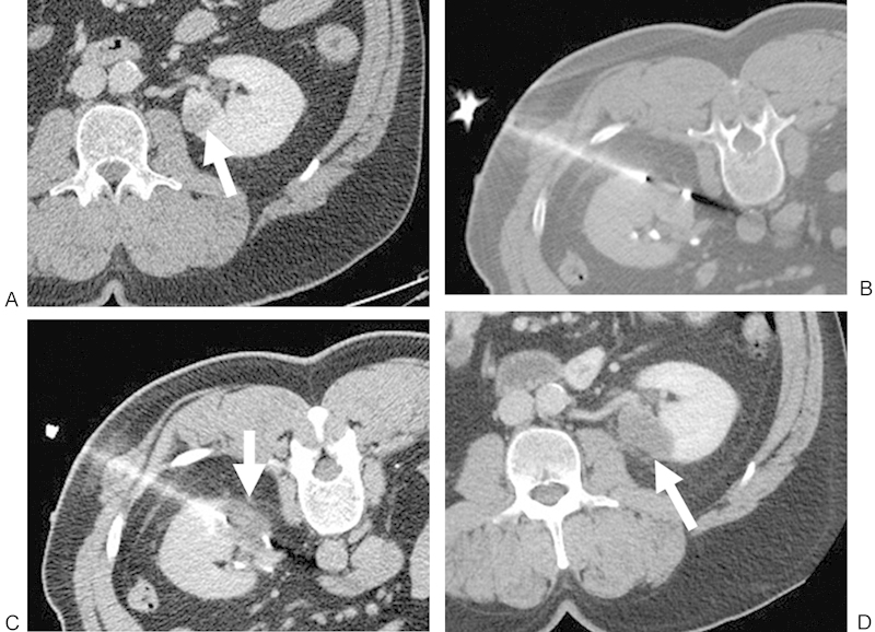
An increasing number of T1a renal cell carcinomas are being diagnosed in recent years, in part due to incidental detection from the increased use of cross-sectional imaging. Although partial nephrectomy is still considered the primary treatment for these small renal masses, percutaneous ablation is now being performed as a standard therapeutic, nephron-sparing approach in patients who are poor surgical candidates. Clinical studies to date have demonstrated that percutaneous ablation is an effective therapy with acceptable outcomes and low risk in the appropriate clinical settings. This article will review various clinical aspects regarding the percutaneous ablation of small renal masses, including patient selection, preprocedural preparations, and the procedural considerations of commonly employed ablative technologies. Specific techniques such as radiofrequency ablation, cryoablation, microwave ablation, irreversible electroporation, and high-intensity focused ultrasound will be addressed in detail. In addition, the technical and oncologic outcomes of percutaneous ablation will be discussed and referenced to that of partial nephrectomy.

Keywords: cryoablation; interventional radiology; percutaneous ablation; radiofrequency ablation; renal mass.


Figures

Figure 1
Figure 1
(A) Contrast-enhanced CT of a 56-year-old man shows a 1.9-cm RCC in the right lower pole (white arrow). The patient was referred for RF because of a complex cardiac history. (B) Noncontrast CT with the patient right-side/ipsilateral-side down demonstrating a posterior approach with the RF probe within the lesion. (C) Noncontrast postablation CT demonstrates areas of increased attenuation (white arrow) in the ablation zone, consistent with blood products, in addition to expected postprocedural changes adjacent to the kidney. This is a common postablation appearance. (D) Contrast-enhanced CT performed approximately 1 month after ablation shows no evidence of enhancement in the ablation zone (white arrow), consistent with a completely treated lesion. CT, computed tomography; RCC, renal cell carcinoma; RF, radiofrequency.
Figure 2
Figure 2
(A) Contrast-enhanced CT of an 83-year-old man shows a 2.3-cm RCC in the interpolar region of the right kidney that extends toward the renal sinus fat (white arrow). The patient was referred for RF because of an overall poor functional status. (B) Noncontrast CT with the patient right-side/ipsilateral-side up demonstrating a lateral approach with the RF probe within the lesion. (C) Noncontrast postablation CT demonstrates areas of increased attenuation (white arrow) in the ablation zone, consistent with blood products, in addition to expected postprocedural changes adjacent to the kidney. This is a common postablation appearance. (D) Contrast-enhanced CT performed approximately 1 month after ablation shows no evidence of enhancement in the ablation zone (white arrow), consistent with a completely treated lesion. CT, computed tomography; RCC, renal cell carcinoma; RF, radiofrequency.
Figure 3
Figure 3
(A) Contrast-enhanced CT of a 55-year-old man shows a 1.8-cm RCC in the lower pole of the left kidney (white arrow). The patient was referred for cryoablation of the left renal lesion because of prior right nephrectomy for RCC. (B) Noncontrast CT with the patient in prone position demonstrating a posterolateral approach with the cryoprobe within the lesion. (C) Noncontrast CT demonstrates the low-attenuation “iceball” in the ablation zone after a 10-minute freeze (white arrow). (D) Contrast-enhanced CT performed approximately 1 month after ablation shows no evidence of enhancement in the ablation zone (white arrow), consistent with a completely treated lesion. CT, computed tomography; RCC, renal cell carcinoma; RF, radiofrequency.

References

    1. National Cancer Institute. Kidney cancer home page Available at: http://www.cancer.gov/cancertopics/types/kidney. Accessed September 25, 2013
    1. Pantuck A J, Zisman A, Belldegrun A S. The changing natural history of renal cell carcinoma. J Urol. 2001;166(5):1611–1623. - PubMed
    1. Gervais D A, McGovern F J, Arellano R S, McDougal W S, Mueller P R. Radiofrequency ablation of renal cell carcinoma: part 1, indications, results, and role in patient management over a 6-year period and ablation of 100 tumors. AJR Am J Roentgenol. 2005;185(1):64–71. - PubMed
    1. Zagoria R J, Hawkins A D, Clark P E. et al.Percutaneous CT-guided radiofrequency ablation of renal neoplasms: factors influencing success. AJR Am J Roentgenol. 2004;183(1):201–207. - PubMed
    1. Jayson M, Sanders H. Increased incidence of serendipitously discovered renal cell carcinoma. Urology. 1998;51(2):203–205. - PubMed