Review
. 2014 Mar;31(1):42-9.
doi: 10.1055/s-0033-1363842.

Percutaneous ablation for small renal masses-complications

Affiliations
Review

Percutaneous ablation for small renal masses-complications

A Nicholas Kurup. Semin Intervent Radiol. 2014 Mar.

Abstract

Although percutaneous ablation of small renal masses is generally safe, interventional radiologists should be aware of the various complications that may arise from the procedure. Renal hemorrhage is the most common significant complication. Additional less common but serious complications include injury to or stenosis of the ureter or ureteropelvic junction, infection/abscess, sensory or motor nerve injury, pneumothorax, needle tract seeding, and skin burn. Most complications may be treated conservatively or with minimal therapy. Several techniques are available to minimize the risk of these complications, and patients should be appropriately monitored for early detection of complications. In the event of a serious complication, prompt treatment should be provided. This article reviews the most common and most important complications associated with percutaneous ablation of small renal masses.

Keywords: complication; cryoablation; interventional radiology; kidney cancer; radiofrequency ablation.


Figures
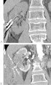
Figure 1
Figure 1
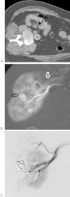
Active extravasation along cryoprobe tracts. (A) Coronal reformatted unenhanced CT during cryoablation shows two of three cryoprobes placed in a 3.7-cm central mass in the right mid kidney of a 72-year-old man. (B) Coronal reformatted contrast-enhanced CT shows active contrast extravasation along the probe tracts (arrows). The patient remained hemodynamically stable, despite a 3.1-g/dL drop in hemoglobin. No expansion of the initially large hematoma was demonstrated on two serial CT scans at 20 and 90 minutes postablation. No transfusion or embolization was performed. CT, computed tomography.
Figure 2
Figure 2
Renal arterial pseudoaneurysm and arteriovenous fistula following radiofrequency ablation. (A) CT shows an electrode in a right renal mass in a 70-year-old woman. (B) DSA image of the right kidney shows a pseudoaneurysm in the right renal hilum (black arrow) as well as early filling of the right renal vein (white arrow). (C) DSA image showing control of the vascular injury postembolization. CT, computed tomography; DSA, digital subtraction angiographic.
Figure 3
Figure 3
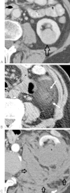
Hemorrhage from ice ball fracture. (A) Contrast-enhanced CT before cryoablation shows a central 4 cm left renal mass (arrow) in an 84-year-old woman. (B) During cryoablation, a fracture (arrows) developed within the ice ball, observed early in the second freeze cycle. (C) As expected, following the cryoablation, a large retroperitoneal hemorrhage developed (arrows). The patient remained hemodynamically stable, despite a hemoglobin drop of 2 g/dL, and did not require treatment. CT, computed tomography.
Figure 4
Figure 4
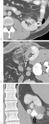
UPJ stricture following renal RFA. (A) Contrast-enhanced CT shows a 1.5-cm mass in the left mid kidney (arrow) of a 46-year-old man. Percutaneous RFA was proposed due to significant medical comorbidities and history of left partial nephrectomy for RCC. (B) Contrast-enhanced CT immediately following RFA shows the ablation zone encompassing the proximal ureter at the UPJ (arrow). (C) Coronal delayed contrast-enhanced CT 3 months later shows a dilated left intrarenal collecting system (arrow) due to UPJ stricture. This kidney eventually became atrophic and nonfunctional. CT, computed tomography; RCC, renal cell carcinoma; RFA, radiofrequency ablation; UPJ, Ureteropelvic junction.
Figure 5
Figure 5
Urine leak after renal radiofrequency ablation. (A) Contrast-enhanced CT shows a 1.7-cm mass in the left mid kidney (arrow) in a 74-year-old woman status postright partial nephrectomy for RCC. (B) Unenhanced CT shows an RFA electrode in the tumor with tip at the renal sinus fat. A small catheter (arrow) is also in place for hydrodisplacement of the adjacent colon. (C) Delayed contrast-enhanced CT immediately following RFA shows urine leaking along the electrode tract (arrows). Due to its persistence on delayed imaging the following day, the urine leak was treated with a ureteral stent for 6 weeks. CT, computed tomography; RCC, renal cell carcinoma; RFA, radiofrequency ablation.
Figure 6
Figure 6
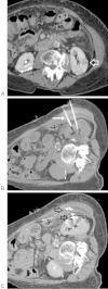
Abscess due to bowel perforation. (A) CT shows a 3.2-cm mass in the left mid kidney (arrow) in an 81-year-old man. (B) CT during cryoablation shows the ice ball (arrows) to encroach upon the descending colon. (C) CT immediately postablation shows complete ablation of the mass. (D) CT performed 2 months later shows a large abscess in the left retroperitoneum and chest wall (arrows), communicating with the ablation zone, presumably related to colonic perforation. CT, computed tomography.
Figure 7
Figure 7
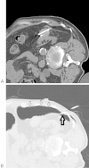
Pneumothorax during renal cryoablation. (A) CT image shows two cryoprobes within an exophytic left renal mass in an 82-year-old man. (B) CT image from a more superior level shows the cryoprobes crossing the pleural space (black arrow) with a moderate-sized pneumothorax (small white arrows). This was resolved with overnight catheter drainage. CT, computed tomography.
Figure 8
Figure 8
Tumor seeding following cryoablation. (A) CT image shows multiple cryoprobes entering through the left anterolateral abdominal wall and an ice ball (arrows) encasing a 3.5-cm left renal cell carcinoma in an 81-year-old man. (B) Postgadolinium MR image shows an enhancing 2-cm nodule (arrow) in the abdominal wall at the site of prior cryoprobe placement. Biopsy confirmed metastatic RCC, and the nodule was then successfully treated with cryoablation. CT, computed tomography; MR, magnetic resonance; RCC, renal cell carcinoma.

References

    1. Atwell T D, Carter R E, Schmit G D. et al.Complications following 573 percutaneous renal radiofrequency and cryoablation procedures. J Vasc Interv Radiol. 2012;23(1):48–54. - PubMed
    1. Dindo D, Demartines N, Clavien P A. Classification of surgical complications: a new proposal with evaluation in a cohort of 6336 patients and results of a survey. Ann Surg. 2004;240(2):205–213. - PMC - PubMed
    1. Morgan M, Smith N, Thomas K, Murphy D G. Is Clavien the new standard for reporting urological complications? BJU Int. 2009;104(4):434–436. - PubMed
    1. Common Terminology Criteria for Adverse Events (CTCAE) Version 4.0. In: U.S. Department of Health and Human Services
    1. Sacks D, McClenny T E, Cardella J F, Lewis C A. Society of Interventional Radiology clinical practice guidelines. J Vasc Interv Radiol. 2003;14(9 Pt 2):S199–S202. - PubMed