. 2014 May;78(4):541-54.
doi: 10.1111/tpj.12495. Epub 2014 Apr 15.

Recognition of floral homeotic MADS domain transcription factors by a phytoplasmal effector, phyllogen, induces phyllody

Affiliations
Free PMC article

Recognition of floral homeotic MADS domain transcription factors by a phytoplasmal effector, phyllogen, induces phyllody

Kensaku Maejima et al. Plant J. 2014 May.
Free PMC article

Abstract

Plant pathogens alter the course of plant developmental processes, resulting in abnormal morphology in infected host plants. Phytoplasmas are unique plant-pathogenic bacteria that transform plant floral organs into leaf-like structures and cause the emergence of secondary flowers. These distinctive symptoms have attracted considerable interest for many years. Here, we revealed the molecular mechanisms of the floral symptoms by focusing on a phytoplasma-secreted protein, PHYL1, which induces morphological changes in flowers that are similar to those seen in phytoplasma-infected plants. PHYL1 is a homolog of the phytoplasmal effector SAP54 that also alters floral development. Using yeast two-hybrid and in planta transient co-expression assays, we found that PHYL1 interacts with and degrades the floral homeotic MADS domain proteins SEPALLATA3 (SEP3), APETALA1 (AP1) and CAULIFLOWER (CAL). This degradation of MADS domain proteins was dependent on the ubiquitin-proteasome pathway. The expression of floral development genes downstream of SEP3 and AP1 was disrupted in 35S::PHYL1 transgenic plants. PHYL1 was genetically and functionally conserved among other phytoplasma strains and species. We designate PHYL1, SAP54 and their homologs as members of the phyllody-inducing gene family of 'phyllogens'.

Keywords: Arabidopsis; MADS domain proteins; floral development; floral quartet model; phyllody; phytoplasma.


Figures

Figure 1
Figure 1
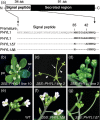
The N–terminal eighth amino acid residue of PHYL1 is required for induction of phyllody-like phenotypes in plants.(a) Schematic representation of PHYL1 and its deletion mutants.(b, c) Floral phenotypes of 35S::PHYL1 transgenic plants.(b) Phyllody-like phenotype, consisting of leaf-like sepals (se), virescent petals (pe) and greenish stamens. A shoot elongated from the initial position of the pistil (arrow), producing a secondary flower.(c) Shoots of secondary flowers arose from the initial position of the pistil (arrow) and from the axils of leaf-like sepals (arrowheads).(d–g) Close-up views of inflorescence apices of (d) 35S::PHYL1, (e) a non-transgenic control plant (Col–0), (f) 35S::PHYL1Δ8, with a normal phenotype, and (g) 35S::PHYL1Δ7, with similar morphological changes to those of 35S::PHYL1 lines.
Figure 2
Figure 2
PHYL1, and its N–terminal eight amino acid deletion mutant, interacts with MADS domain proteins SEP3, AP1 and CAL in yeast.The MADS domain proteins SEP3, AP1 and CAL, as well as WUS, fused to the GAL4 activation domain (AD), were expressed in combination with SAP54, PHYL1 and PHYL1Δ8 fused to the GAL4 DNA-binding domain (BD) in yeast strain AH109. As negative controls, each AD-fused host factor was co-expressed with empty BD vector. Yeast cells harboring the AD and BD vectors were cultured to an absorbance of 0.1 at 600 nm. Aliquots (10 μL) of these cells were plated on selective medium lacking leucine and tryptophan (–LW), or lacking leucine, tryptophan and histidine (–LWH). The plates were incubated for 3 days at 30°C.
Figure 3
Figure 3
PHYL1 inhibits MADS domain protein functions.(a) Quantitative real-time RT–PCR analysis of expression levels of genes downstream of SEP3 and AP1 (AP3, PI, SOC1, SVP and AGL24) in wild-type and 35S::PHYL1 transgenic plants. The expression level of the genes in wild-type plants was set as the reference. Error bars represent the standard deviation. Asterisks indicate statistically significant differences between wild-type and 35S::PHYL1 transgenic plants (*P < 0.05, **P < 0.01).(b–d) BiFC assays showing that PHYL1 interferes with the interaction between the MADS domain proteins. Agrobacterium cultures (OD600 = 1.0) expressing the N- and C–terminal YFP-fused proteins and either GUS (from pCAMBIA1301 i GenBark accession number AF234297), PHYL1 or PHYL1Δ8 were mixed at a ratio of 1:1:10. YFP fluorescence was detected 36 h after co-expression of (b) bZIP63–NYF and bZIP63–CYF, (c) NYF–SEP3 and AP1–CYF, and (d) NYF–SEP3 and CYF–AG, with GUS, PHYL1 or PHYL1Δ8. Scale bar = 200 μm.(e) Quantitative analysis of the number of nuclear-localized BiFC signals in (b–d). The number of signals was quantified using a leaf area of 2.4 mm2. Error bars represent the standard deviation. Asterisks indicate statistically significant differences compared with GUS (*P < 0.05, **P < 0.01).
Figure 4
Figure 4
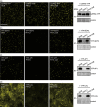
Transient expression of PHYL1 induces degradation of MADS domain proteins.(a–d) Accumulation and subcellular localization of transiently expressed YFP-fused Arabidopsis nuclear protein bZIP63 (a) and YFP-fused MADS domain proteins SEP3, AP1 and CAL (b–d). Agrobacterium cultures (OD600 = 1.0) expressing YFP-fused proteins and either GUS, PHYL1 or PHYL1Δ8 were mixed at a ratio of 1:10, and infiltrated into N. benthamiana leaves. YFP fluorescence was observed 36 h after expression. Scale bar = 200 μm. Accumulation of YFP-fused proteins was evaluated by immunoblotting using an anti-GFP antibody (a–d, right).
Figure 5
Figure 5
PHYL1-induced degradation of MADS domain proteins is mediated by the ubiquitin–proteasome pathway.(a) Confocal images of N. benthamiana leaf epidermis cells transiently expressing YFP–SEP3 and PHYL1 following infiltration with dimethylsulfoxide (DMSO) (left) or 20 μm of the proteasome inhibitor clasto-lactacystin β–lactone (right). Agrobacterium cultures expressing YFP–SEP3 and PHYL1 were mixed at a ratio of 1:1, and infiltrated into N. benthamiana leaves. DMSO or clasto-lactacystin β–lactone were infiltrated 1 day after co-infiltration of YFP–SEP3 and PHYL1, and YFP fluorescence was observed 12 h after clasto-lactacystin β–lactone or DMSO infiltration. Scale bar = 200 μm.(b) Immunoprecipitation of bZIP63–YFP or YFP–SEP3 using an anti-GFP antibody, and detection of ubiquitinated proteins using an anti-ubiquitin antibody. Agrobacterium cultures expressing YFP-fused proteins and either PHYL1 or buffer were mixed at a ratio of 1:1 and infiltrated into N. benthamiana leaves. Thirty-six hours after expression, total proteins were extracted from N. benthamiana leaves transiently co-expressing bZIP63–YFP and PHYL1 (left lane), YFP–SEP3 alone (middle lane) or YFP–SEP3 and PHYL1 (right lane), and immunoprecipitation (IP) was performed using an anti-GFP antibody. Total proteins (upper panel) and immunoprecipitates (middle and lower panels) were subjected to SDS–PAGE and immunoblotting (IB) using anti-GFP antibody (upper and middle panels) and anti-ubiquitin antibody (lower panel), as indicated.
Figure 6
Figure 6
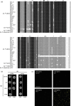
Identification and functional characterization of PHYL1 homologs.(a) Amino acid alignment of PHYL1 homologs. The full strain names and GenBank accession numbers of PHYL1 homologs are listed in Table S2. Previously reported homologs are indicated by dagger(s): one dagger, Jomantiene et al. (2007); two daggers, MacLean et al. (2011); three daggers, Saccardo et al. (2012). The arrowhead indicates the putative signal peptide cleavage site predicted by SignalP software (http://www.cbs.dtu.dk/services/SignalP/). All PHYL1 homologs encode putative secretory peptides similar to PHYL1, of which the C–terminal 91 residues are secreted to the host cytoplasm. The background indicates the percentage amino acid similarity: black, 100%; dark gray, 80%; light gray, 60%.(b) Yeast two-hybrid assay showing the interaction between SEP3 and PHYL1 homologs. A MADS domain protein SEP3 fused to the GAL4 activation domain (AD) or empty AD vector was expressed in combination with PHYL1 homologs CA–76, CP, PEY and PYR fused to the GAL4 DNA-binding domain (BD) in yeast strain AH109. As negative controls, each BD-fused host factor was co-expressed with empty AD vector. Experimental details are as described in the legend to Figure2.(c) Transient co-expression assay showing that PHYL1 homologs CA–76, CP and PEY induced degradation of YFP–SEP3. YFP–SEP3 was co-expressed with the PHYL1 homologs, and YFP fluorescence was observed 36 h after expression. Experimental details are as described in the legend to Figure4.
Figure 7
Figure 7
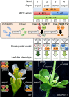
Model of induction of phyllody-like symptoms by a phytoplasma effector ‘phyllogen’.

References

    1. Abramovitch RB, Anderson JC, Martin GB. Bacterial elicitation and evasion of plant innate immunity. Nat. Rev. Mol. Cell Biol. 2006;7:601–611. - PMC - PubMed
    1. Alvarez-Buylla ER, Benítez M, Corvera-Poiré A, et al. Flower development. Arabidopsis Book. 2010;8:e0127. - PMC - PubMed
    1. Bertaccini A, Duduk B. Phytoplasma and phytoplasma diseases: a review of recent research. Phytopathol. Mediterr. 2009;48:355–378.
    1. Bowman JL, Smyth DR, Meyerowitz EM. Genes directing flower development in Arabidopsis. Plant Cell. 1989;1:37–52. - PMC - PubMed
    1. Bowman JL, Alvarez J, Weigel D, Meyerowitz EM, Smyth DR. Control of flower development in Arabidopsis thaliana by APETALA1 and interacting genes. Development. 1993;119:721–743.

Publication types

MeSH terms