. 2014 Mar 6;9(1):108.
doi: 10.1186/1556-276X-9-108.

Cellular distribution and cytotoxicity of graphene quantum dots with different functional groups

Affiliations

Cellular distribution and cytotoxicity of graphene quantum dots with different functional groups

Xiaochan Yuan et al. Nanoscale Res Lett. .

Abstract

Graphene quantum dots (GQDs) have been developed as promising optical probes for bioimaging due to their excellent photoluminescent properties. Additionally, the fluorescence spectrum and quantum yield of GQDs are highly dependent on the surface functional groups on the carbon sheets. However, the distribution and cytotoxicity of GQDs functionalized with different chemical groups have not been specifically investigated. Herein, the cytotoxicity of three kinds of GQDs with different modified groups (NH2, COOH, and CO-N (CH3)2, respectively) in human A549 lung carcinoma cells and human neural glioma C6 cells was investigated using thiazoyl blue colorimetric (MTT) assay and trypan blue assay. The cellular apoptosis or necrosis was then evaluated by flow cytometry analysis. It was demonstrated that the three modified GQDs showed good biocompatibility even when the concentration reached 200 μg/mL. The Raman spectra of cells treated with GQDs with different functional groups also showed no distinct changes, affording molecular level evidence for the biocompatibility of the three kinds of GQDs. The cellular distribution of the three modified GQDs was observed using a fluorescence microscope. The data revealed that GQDs randomly dispersed in the cytoplasm but not diffused into nucleus. Therefore, GQDs with different functional groups have low cytotoxicity and excellent biocompatibility regardless of chemical modification, offering good prospects for bioimaging and other biomedical applications.


Figures

Figure 1
Figure 1
UV–Vis absorption spectra and fluorescence spectra of three kinds of GQDs. (a) The UV–Vis absorption spectra of three kinds of GQDs. (b) The fluorescence spectra of aGQDs excited from 320 to 580 nm. (c) The fluorescence spectra of cGQDs independent on the excitation wavelength. (d) The fluorescence spectra of dGQDs.
Figure 2
Figure 2
TEM images of three modified GQDs deposited on copper grids. (a) The TEM image of aGQDs. (b) Diameter distribution of the cGQDs. (c) The TEM image of dGQDs.
Figure 3
Figure 3
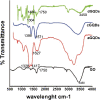
FTIR spectra of the GQDs. The FTIR spectra of three modified GQDs and GO.
Figure 4
Figure 4
Representative fluorescence microscope images of cells. (a) Fluorescence image describing control cells. (b) Cells treated with 50 μg/mL of aGQDs for 12 h. (c) Cells exposed to 50 μg/mL of cGQDs for 12 h. (d) Cells after the treatment of 50 μg/mL of dGQDs for 12 h. Magnification, ×20.
Figure 5
Figure 5
The MTT (% of control) evaluated after exposed to three kinds of GQDs for 24 h. (a) MTT (% of control) of A549 cells after exposed to different concentrations of three kinds of GQDs. (b) MTT (% of control) of C6 after the exposure to three kinds of GQDs at different concentrations. Asterisk indicated p < 0.05 and double asterisk represented p < 0.01.
Figure 6
Figure 6
The influence of GQDs with different functional groups on the mortality of cells. (a) Cell mortality of A549 cells after treated with different concentrations of three GQDs. (b) Cell mortality after exposed to different concentrations of three kinds of GQDs evaluated in C6 cell line. Asterisk indicated p < 0.05 and double asterisk represented p < 0.01.
Figure 7
Figure 7
Representative FACS images and the statistical results of cell apoptosis rate and necrosis rate. After exposed to 200 μg/mL of the three kinds of GQDs. (a) Statistical results of cell necrosis. (b) Statistical results of cell apoptosis.
Figure 8
Figure 8
Raman spectra of cells. (a) Mean Raman spectra of A549 cells before and after exposure to 200 μg/mL of GQDs. (b) Average Raman spectra of C6 cells before and after treated with GQDs at the concentration of 200 μg/mL. Excitation wavelength, 785 nm.

References

    1. Shao L, Gao Y, Yan F. Semiconductor quantum dots for biomedicial applications. Sensors. 2011;9:11736–11751. doi: 10.3390/s111211736. - DOI - PMC - PubMed
    1. Valizadeh A, Mikaeili H, Samiei M, Farkhani S, Zarghami N, Kouhi M, Akbarzadeh A, Davaran S. Quantum dots: synthesis, bioapplications, and toxicity. Nanoscale Res Lett. 2012;9:480. doi: 10.1186/1556-276X-7-480. - DOI - PMC - PubMed
    1. Gomes S, Vieira C, Almeida D, Santos-Mallet J, Menna-Barreto R, Cesar C, Feder D. CdTe and CdSe quantum dots cytotoxicity: a comparative study on microorganisms. Sensors. 2011;9:11664–11678. doi: 10.3390/s111211664. - DOI - PMC - PubMed
    1. Liu L, Miao Q, Liang G. Quantum dots as multifunctional materials for tumor imaging and therapy. Materials. 2013;9:483–499. doi: 10.3390/ma6020483. - DOI - PMC - PubMed
    1. Qu G, Wang X, Wang Z, Liu S, Jiang G. Cytotoxicity of quantum dots and graphene oxide to erythroid cells and macrophages. Nanoscale Res Lett. 2013;9:198. doi: 10.1186/1556-276X-8-198. - DOI - PMC - PubMed

LinkOut - more resources