. 2014 Aug;19(7):1024-34.
doi: 10.1177/1087057114525853. Epub 2014 Mar 5.

Enzymatic Characterization of ER Stress-Dependent Kinase, PERK, and Development of a High-Throughput Assay for Identification of PERK Inhibitors

Affiliations

Enzymatic Characterization of ER Stress-Dependent Kinase, PERK, and Development of a High-Throughput Assay for Identification of PERK Inhibitors

Dariusz Pytel et al. J Biomol Screen. 2014 Aug.

Abstract

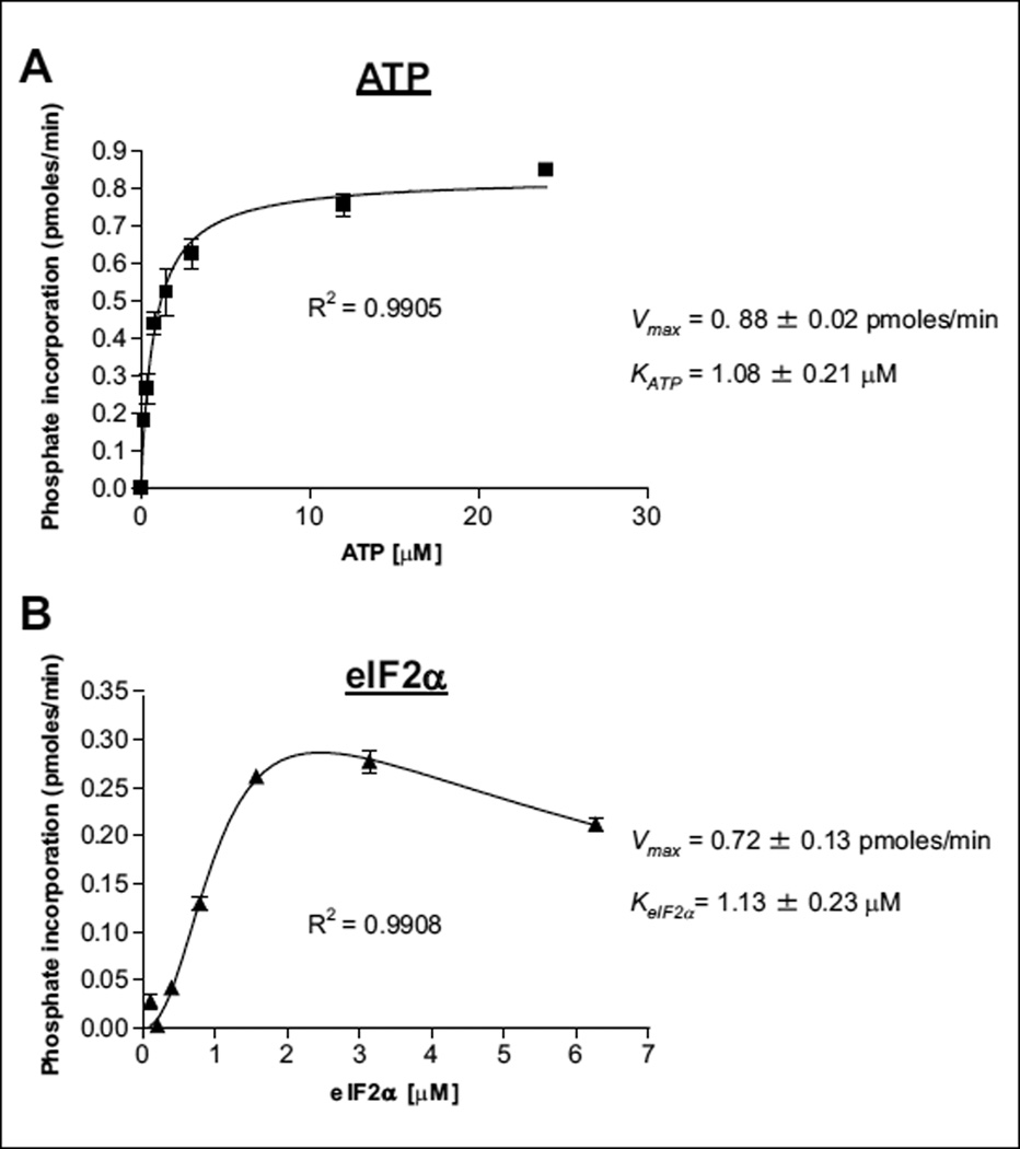
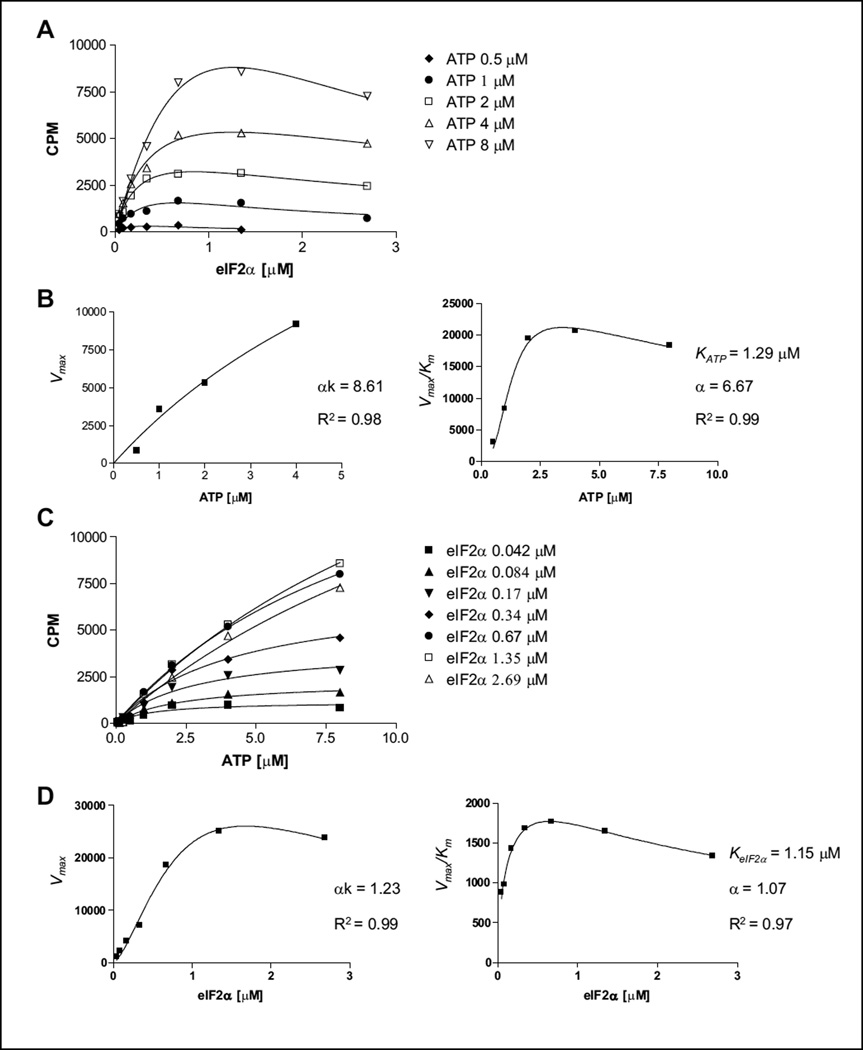
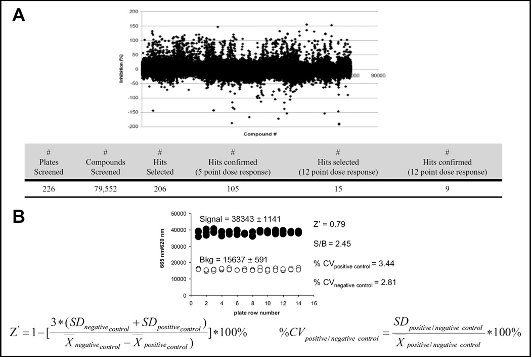
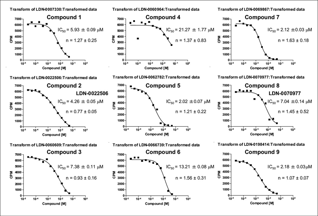
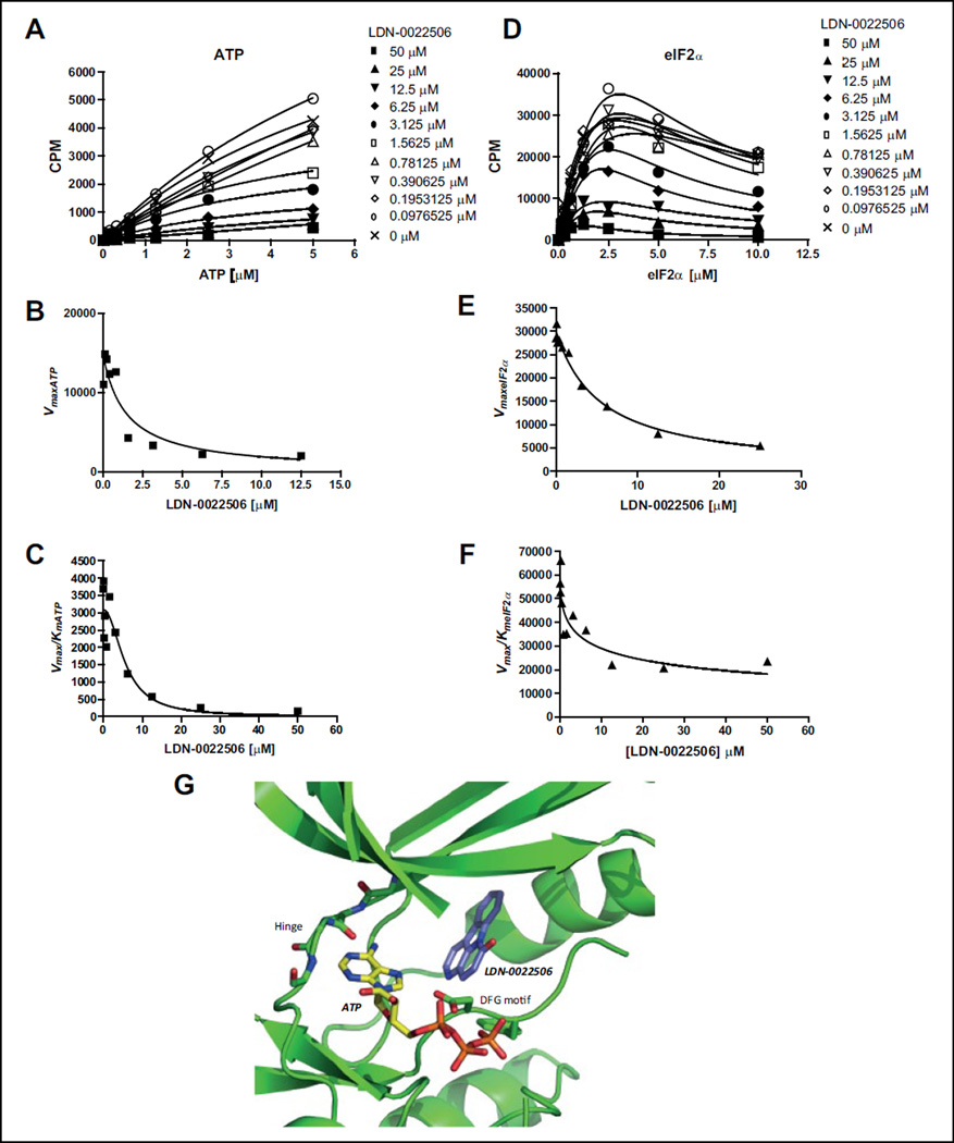
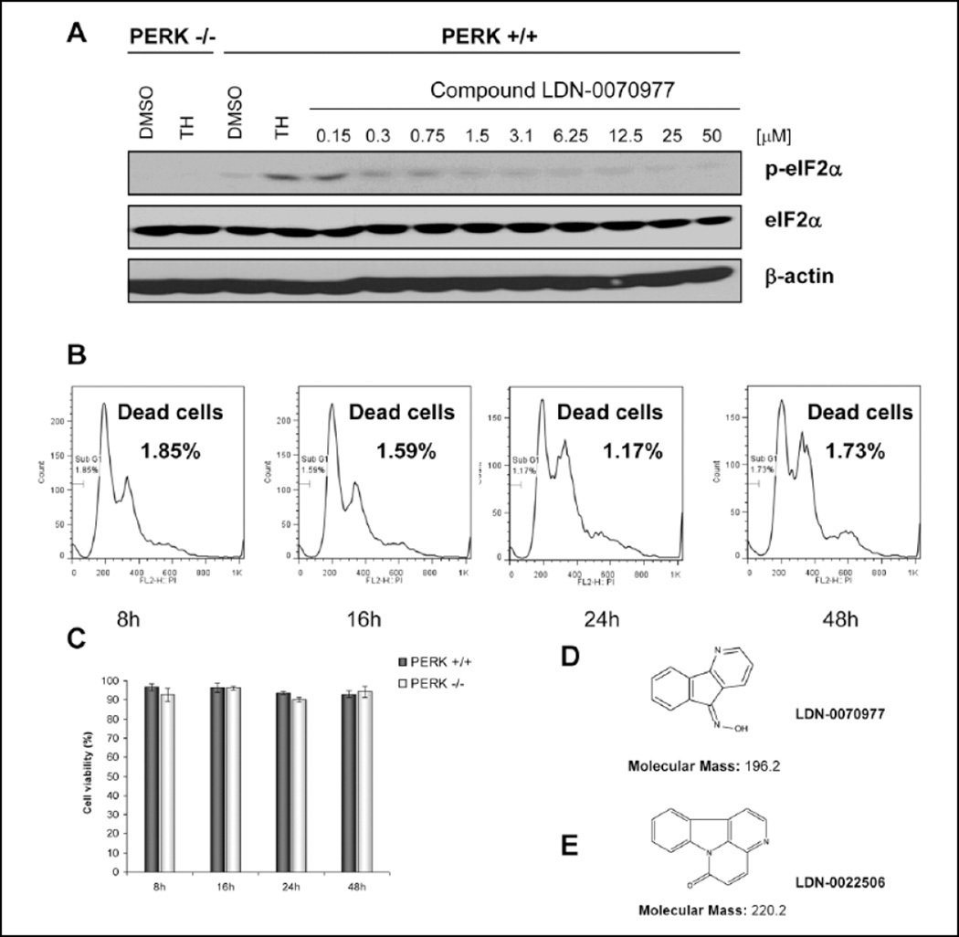
PERK is serine/threonine kinase localized to the endoplasmic reticulum (ER) membrane. PERK is activated and contributes to cell survival in response to a variety of physiological stresses that affect protein quality control in the ER, such as hypoxia, glucose depravation, increased lipid biosynthesis, and increased protein translation. Pro-survival functions of PERK are triggered by such stresses, suggesting that development of small-molecule inhibitors of PERK may be efficacious in a variety of disease scenarios. Hence, we have conducted a detailed enzymatic characterization of the PERK kinase to develop a high-throughput-screening assay (HTS) that will permit the identification of small-molecule PERK inhibitors. In addition to establishing the K(m) of PERK for both its primary substrate, eIF2α, and for adenosine triphosphate, further mechanistic studies revealed that PERK targets its substrate via either a random/steady-state ordered mechanism. For HTS, we developed a time-resolved fluorescence resonance energy transfer-based assay that yielded a robust Z' factor and percent coefficient of variation value, enabling the successful screening of 79,552 compounds. This approach yielded one compound that exhibited good in vitro and cellular activity. These results demonstrate the validity of this screen and represent starting points for drug discovery efforts.

Keywords: HTS; PERK; small-molecule inhibitors.


Conflict of interest statement

Declaration of Conflicting Interests

The authors declared no potential conflicts of interest with respect to the research, authorship, and/or publication of this article.

Figures

Figure 1
Figure 1
Kinetic characterization of PERK enzyme substrates. Initial velocity measurements were used for the (A) Km determination for adenosine triphosphate (ATP) and (B) Km determination for eIF2α by the radiometric kinase assay. In total, 8 nM PERK and 2 µM eIF2α was used for the ATP titration (A), 8-nM PERK, and 1 µM ATP was used for the eIF2α titration (B).
Figure 2
Figure 2
Kinetic mechnism studies for PERK toward adenosine triphosphate (ATP) and eIF2α substrates. (A) Titration of ATP in the range of 0.5 to 8 µM versus an eIF2a concentration of 0.04 to 3 µM. From each ATP concentration plot, Vmax values of each reaction were calculated. (B) Determination of αk = 8.61 and α = 6.67 values. Based on the curve fit, we demonstrate that PERK kinase follows a random mechanism toward the ATP substrate. (C) Titration of eIF2α in the range of 0.04 to 3 µM versus an ATP concentration of 0.5 to 8 µM. From each eIF2α concentration plot, Vmax values of each reaction were calculated. (D) Determination of αk = 1.23 and α = 1.07 values. Based on the curve fit, we demonstrate that PERK kinase follows a random or steady-state ordered mechanism toward the eIF2α substrate. Experiments were repeated a minimum of three times. One representative experiment is shown.
Figure 3
Figure 3
High-throughput screen of a small-molecule compounds library. (A) Graph representing % of PERK inhibition in the fluorescence resonance energy transfer (FRET)–based kinase assay and high-throughput screening results. (B) Statistics for the high-throughput screen were determined (Z′; % coefficient of variation [CV] of positive and negative controls and signal-to-background [S/B] ratio). The background was run in the absence of PERK.
Figure 4
Figure 4
Evaluation of compounds by radiometric assay. Compounds were evaluated at 1 µM eIF2α, 1 µM adenosine triphosphate (ATP), and 8 nM PERK. The IC50 for each compound was averaged from the three independent experiments, and one representative experiment is shown. The sigmoidal dose-response (variable slop) equation was used to obtain curve fits.
Figure 5
Figure 5
Inhibition mechanism study of LDN-0022506. (A) Plot of initial velocities vs [ATP] at different concentrations of LDN-0022506, all at a fixed eIF2α concentration of 1 µM. (B, C) LDN-0022506 concentration dependencies of (Vmax)ATP and (Vmax/Km)ATP apparent values derived from analysis of the data of panel A. (D) Plot of initial velocities vs [eIF2α] at different concentrations of LDN-0022506, all at a fixed ATP concentration of 1 µM. (E, F) LDN-0022506 concentration dependencies of (Vmax)eIF2α and (Vmax/Km)eIF2α apparent values derived from analysis of the data of panel D. Experiments were repeated a minimum of three times. One representative experiment is shown. (G) ATP molecule and LDN-0020506 were docked into the crystal structure of PERK kinase domain (4G31). Docking calculations were carried out using induced fit docking with Glide and Prime as part of the Schrodinger First Discovery Suite.
Figure 6
Figure 6
Compound LDN-0070977 inhibits PERK phosphorylation with no toxicity to the cells. (A) Mouse embryonic fibroblast (MEF) cells were pretreated with LDN-0070977 inhibitor in the range of 0.15 to 50 µM for 1 h; next cells were treated with 500 nM thapsigargin (TH) to activate PERK. PERK−/− MEFs were used as a control. Cells were lysed and analyzed by Western blot. (B, C) Compound toxicity was measured in 3T3 cells by propidium iodide (B) and trypan blue staining (C). (D) Structure of LDN-0070977 compound. (E) Structure of LDN-0022506 compound.

References

    1. Shi Y, Vattem KM, Sood R, et al. Identification and Characterization of Pancreatic Eukaryotic Initiation Factor 2 Alpha-Subunit Kinase, PEK, Involved in Translational Control. Mol. Cell Biol. 1998;18:7499–7509. - PMC - PubMed
    1. Wang XZ, Harding HP, Zhang Y, et al. Cloning of Mammalian Ire1 Reveals Diversity in the ER Stress Responses. Embo J. 1998;17:5708–5717. - PMC - PubMed
    1. Haze K, Yoshida H, Yanagi H, et al. Mammalian Transcription Factor ATF6 Is Synthesized as a Transmembrane Protein and Activated by Proteolysis in Response to Endoplasmic Reticulum Stress. Mol. Biol. Cell. 1999;10:3787–3799. - PMC - PubMed
    1. Yamamoto K, Sato T, Matsui T, et al. Transcriptional Induction of Mammalian ER Quality Control Proteins Is Mediated by Single or Combined Action of ATF6alpha and XBP1. Dev. Cell. 2007;13:365–376. - PubMed
    1. Wu J, Rutkowski DT, Dubois M, et al. ATF6alpha Optimizes Long-Term Endoplasmic Reticulum Function to Protect Cells from Chronic Stress. Dev. Cell. 2007;13:351–364. - PubMed

Publication types