. 2014 Mar 25;82(12):1068-75.
doi: 10.1212/WNL.0000000000000241. Epub 2014 Mar 5.

Role of the phosphoinositide phosphatase FIG4 gene in familial epilepsy with polymicrogyria

Affiliations

Role of the phosphoinositide phosphatase FIG4 gene in familial epilepsy with polymicrogyria

Stéphanie Baulac et al. Neurology. .

Abstract

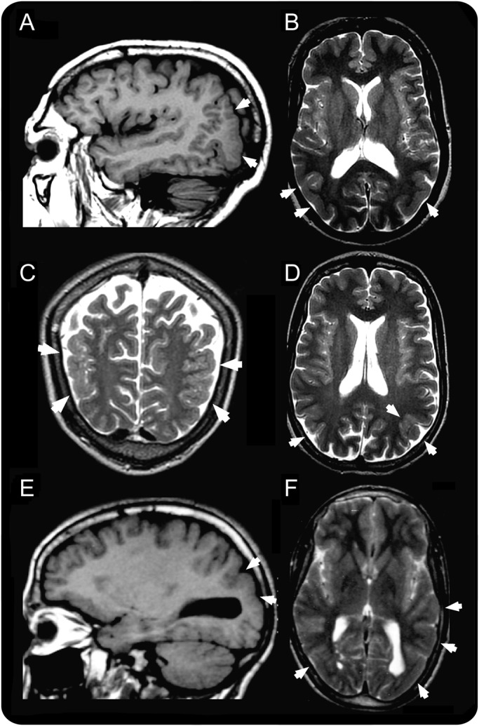
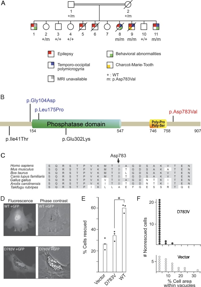
Objective: The aim of this study was to identify the causal gene in a consanguineous Moroccan family with temporo-occipital polymicrogyria, psychiatric manifestations, and epilepsy, previously mapped to the 6q16-q22 region.

Methods: We used exome sequencing and analyzed candidate variants in the 6q16-q22 locus, as well as a rescue assay in Fig4-null mouse fibroblasts and immunohistochemistry of Fig4-null mouse brains.

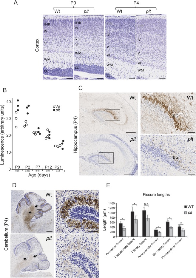
Results: A homozygous missense mutation (p.Asp783Val) in the phosphoinositide phosphatase gene FIG4 was identified. Pathogenicity of the variant was supported by impaired rescue of the enlarged vacuoles in transfected fibroblasts from Fig4-deficient mice. Histologic examination of Fig4-null mouse brain revealed neurodevelopmental impairment in the hippocampus, cortex, and cerebellum as well as impaired cerebellar gyration/foliation reminiscent of human cortical malformations.

Conclusions: This study extends the spectrum of phenotypes associated with FIG4 mutations to include cortical malformation associated with seizures and psychiatric manifestations, in addition to the previously described Charcot-Marie-Tooth disease type 4J and Yunis-Varón syndrome.


Figures

Figure 1
Figure 1. Brain MRI
(A, B) Patient V.8, (C, D) patient V.9, and (E, F) patient V.11. Images A and E are T1-weighted sagittal sections; B, D, and F are T2-weighted axial sections; and C is a T2-weighted coronal section. The areas of abnormal cortical development in the temporo-occipital areas are indicated by the white arrows and are consistent in all 3 patients with a combination of increased cortical thickness, smooth cortex, overfolding, and microgyri.
Figure 2
Figure 2. Identification of FIG4 mutation
(A) Pedigree of the family with segregation of the FIG4 variant. Only generations IV and V are shown. (B) FIG4 is a multidomain protein with a protein interaction domain at the N-terminus, a SAC phosphatase domain in the central region, and poly-Pro and poly-Ser domains within the C-terminal. The mutation identified in this study is indicated in red, and missense mutations previously reported in Charcot-Marie-Tooth disease type 4J and Yunis-Varón syndrome are indicated in black and blue, respectively. (C) Multiple protein alignment showing conservation of the aspartate (D) residue in position 783 in orthologs of FIG4 across species. (D) Phenotype rescue in Fig4-null fibroblasts of plt mice. Mutant or wild-type (WT) Fig4 cDNA was cotransfected with GFP into Fig4-null fibroblasts. The large intracytoplasmic vesicles characteristic of Fig4-null fibroblasts were rescued in 60% of cells by the WT cDNA (top panels) but in only 33% of cells by the D783V mutant cDNA (bottom panels). (E) Percent of transfected (GFP-positive) fibroblasts that lack vacuoles. The results of 3 independent experiments are indicated. *p < 0.01. (F) The D783V mutant retains partial activity. In nonrescued cells from panel E, the percentage of cell area covered by vacuoles in cells expressing D783V (3.3% ± 0.6%, n = 24) is smaller than that in cells receiving vector only (10.3% ± 1.8%, n = 22) (2-tailed p value <0.001). Transfection of the mutant cDNA does not completely rescue the formation of vacuoles but does reduce the severity of vacuolization. cDNA = complementary DNA; GFP = green fluorescent protein; plt = pale tremor.
Figure 3
Figure 3. Neurodevelopmental anomalies in plt mice
(A) Nissl staining of cortex from plt homozygote and WT littermate at postnatal days P0 and P4. Note presence of swollen neurons in layers IV and V of plt aged P4. Cortical layers are indicated as follows: I–VI, WM (white matter), CC (corpus callosum). (B) Caspase 3/7 activity was measured in brain homogenates from 3 animals of each genotype at the indicated postnatal age. An unpaired Student t test was used to generate p values at each age. (C) Weak calbindin immunoreactivity in hippocampus of plt mouse compared with WT littermates. Higher magnification of dorsal DG (boxed regions) shows many granule cells with a well-developed dendrite tree in the WT brain, while in the plt, there is weak staining of the perikaryon and no immunostaining of dendrite trees. (D) Very weak calbindin immunoreactivity in cerebellar cortex and deep nucleus (arrows) in plt mouse compared with WT littermate at P4. Boxed areas are focused on posterolateral fissures. Scale bars: A, 100 μm; C, 300 μm (boxed area 50 μm); D, 250 μm (boxed area 50 μm). (E) Histogram representing the fissure lengths in the cerebellum at P4 (n = 5). Significant differences between lengths of fissures were observed between WT and plt. *p < 0.05; **p < 0.01. DG = dentate gyrus; n.s. = not significant; plt = pale tremor; WT = wild-type.

References

    1. Barkovich AJ, Guerrini R, Kuzniecky RI, Jackson GD, Dobyns WB. A developmental and genetic classification for malformations of cortical development: update 2012. Brain 2012;135:1348–1369 - PMC - PubMed
    1. Piao X, Hill RS, Bodell A, et al. G protein-coupled receptor-dependent development of human frontal cortex. Science 2004;303:2033–2036 - PubMed
    1. Valence S, Poirier K, Lebrun N, et al. Homozygous truncating mutation of the KBP gene, encoding a KIF1B-binding protein, in a familial case of fetal polymicrogyria. Neurogenetics 2013;14:215–224 - PubMed
    1. Morris-Rosendahl DJ, Segel R, Born AP, et al. New RAB3GAP1 mutations in patients with Warburg Micro syndrome from different ethnic backgrounds and a possible founder effect in the Danish. Eur J Hum Genet 2010;18:1100–1106 - PMC - PubMed
    1. Borck G, Wunram H, Steiert A, et al. A homozygous RAB3GAP2 mutation causes Warburg Micro syndrome. Hum Genet 2011;129:45–50 - PubMed

Publication types

MeSH terms