Comparative Study
. 2014 Mar;70(Pt 3):708-19.
doi: 10.1107/S1399004713032446. Epub 2014 Feb 15.

Role of κ→λ light-chain constant-domain switch in the structure and functionality of A17 reactibody

Affiliations
Comparative Study

Role of κ→λ light-chain constant-domain switch in the structure and functionality of A17 reactibody

Natalia Ponomarenko et al. Acta Crystallogr D Biol Crystallogr. 2014 Mar.

Abstract

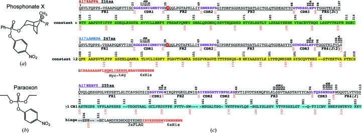
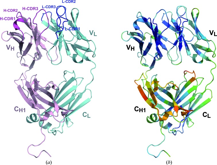
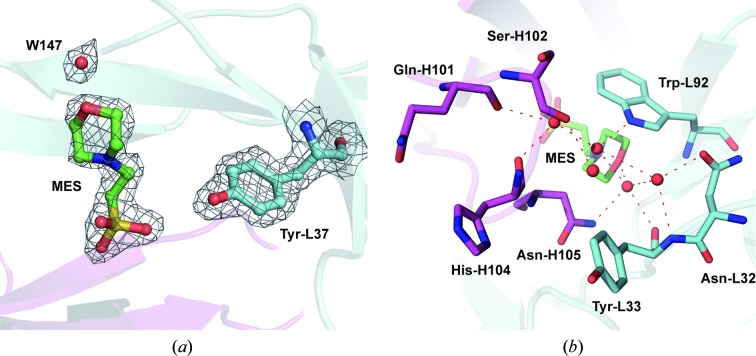
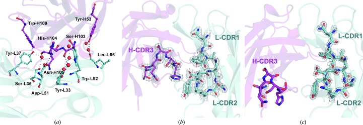
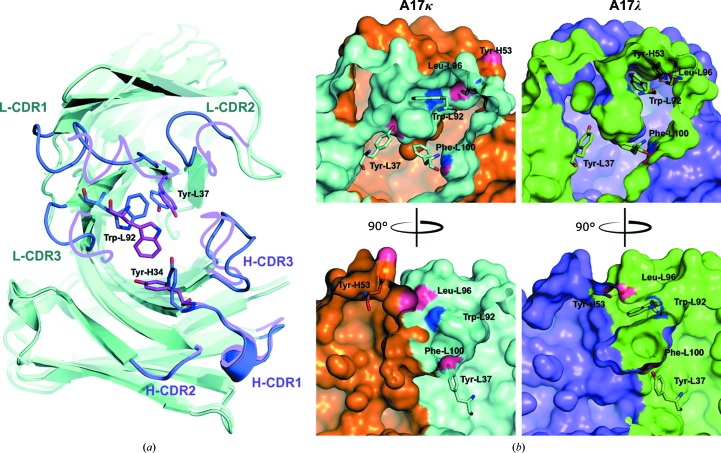
The engineering of catalytic function in antibodies requires precise information on their structure. Here, results are presented that show how the antibody domain structure affects its functionality. The previously designed organophosphate-metabolizing reactibody A17 has been re-engineered by replacing its constant κ light chain by the λ chain (A17λ), and the X-ray structure of A17λ has been determined at 1.95 Å resolution. It was found that compared with A17κ the active centre of A17λ is displaced, stabilized and made more rigid owing to interdomain interactions involving the CDR loops from the VL and VH domains. These VL/VH domains also have lower mobility, as deduced from the atomic displacement parameters of the crystal structure. The antibody elbow angle is decreased to 126° compared with 138° in A17κ. These structural differences account for the subtle changes in catalytic efficiency and thermodynamic parameters determined with two organophosphate ligands, as well as in the affinity for peptide substrates selected from a combinatorial cyclic peptide library, between the A17κ and A17λ variants. The data presented will be of interest and relevance to researchers dealing with the design of antibodies with tailor-made functions.

Keywords: A17 reactibody; antibodies.


Figures

Figure 1
Figure 1
Chemical compounds and antibody chains used in this study. (a) R = H, p-nitrophenyl 8-methyl-8-azabicyclo[3.2.1]octylphenylphosphonate (phosphonate X); R = biotin, biotinylated phosphonate X (BtX). (b) O,O-Diethyl O-(4-nitrophenyl)phosphate (paraoxon). (c) Amino-acid sequences of A17 κ and λ light chains and the heavy chain. Residues are numbered using the Kabat system (superlinear) and sequential numbering (interlinear). Frameworks (FR1–FR4) are underlined; switch residues of the J segment are designated as FR4 according to Kabat; CDRs are coloured magenta; constant domains κCL, λCL and CH1 are coloured green, yellow and blue, respectively.
Figure 2
Figure 2
(a) The overall structure of the A17λ antibody. Heavy (VH/CH1) and light chains are shown in magenta and cyan, respectively. (b) The A17λ structure coloured according to the Cα atomic displacement parameters (ADP), with a colour transition from blue to red indicating increasing ADP values.
Figure 3
Figure 3
(a) 2F oF c electron-density map contoured at the 1.5σ level above the mean (0.4 e Å−3) showing the MES molecule, water 147 and the catalytic Tyr-L36. Heavy and light chains are shown in magenta and cyan, respectively. (b) An extended hydrogen-bonding pattern involving the MES molecule and five intact water molecules located close to the A17λ active centre (red dashed lines).
Figure 4
Figure 4
(a) Interdomain interactions between the CDR loops of A17λ and interactions involving catalytic Tyr-L37. Protein residues involved in these interactions are shown in ball-and-stick representation, water molecules are shown as spheres and hydrogen-bond interactions are shown as blue dashed lines. Here and in (b) and (c), heavy and light chains are shown in magenta and cyan, respectively. Tyr-L33 and Ser-L35 belong to L-CDR1, Asp-L51 to L-­CDR2, Trp-L92 and LeuL96 to L-CDR3, Tyr-H53 to H-CDR2, and Ser-H103, His-H104 and Asn-H105 to H-CDR3. (b) 2F oF c electron-density map contoured at the 2σ level (0.5 e Å−3) above the mean for the CDR loop region of A17λ. (c) 2F oF c electron-density map contoured at the 2σ level (0.5 e Å−3) above the mean for the CDR loop region of A17κ.
Figure 5
Figure 5
(a) Superposition of the A17λ and A17κ variable domains. A17λ CDR loops are shown in magenta and A17κ CDR loops are shown in blue. The flip of the Trp-L92 side chain is indicated. (b) The flipping of Trp-L92 provides the enlargement of the hydrophobic pocket surface (Trp-L92–Phe-L100) and the formation of a kind of lid above the cavity entrance. The light chain of A17κ is shown in cyan and the heavy chain is shown in brown; the light chain of A17λ is shown in green and the heavy chain in shown in magenta.
Figure 6
Figure 6
Structural superposition of the A17κ (blue) and A17λ (magenta) light chains on the VL domain. The Gly-L110 insertion in A17λ accounts for the change in the VL–CL orientation compared with that in A17κ.
Figure 7
Figure 7
Elbow angles in A17 reactibody variants. A17κ-P is a phosphonylated variant.

References

    1. Afonine, P. V., Grosse-Kunstleve, R. W., Echols, N., Headd, J. J., Moriarty, N. W., Mustyakimov, M., Terwilliger, T. C., Urzhumtsev, A., Zwart, P. H. & Adams, P. D. (2012). Acta Cryst. D68, 352–367. - PMC - PubMed
    1. Eddleston, M., Buckley, N. A., Eyer, P. & Dawson, A. H. (2008). Lancet, 371, 597–607. - PMC - PubMed
    1. Ekiert, D. C. et al. (2012). Nature (London), 489, 526–532. - PubMed
    1. Emsley, P. & Cowtan, K. (2004). Acta Cryst. D60, 2126–2132. - PubMed
    1. Evans, P. (2006). Acta Cryst. D62, 72–82. - PubMed

Publication types

MeSH terms

Substances

Associated data