. 2014 Mar;70(Pt 3):744-51.
doi: 10.1107/S139900471303294X. Epub 2014 Feb 15.

Structural evidence for the partially oxidized dipyrromethene and dipyrromethanone forms of the cofactor of porphobilinogen deaminase: structures of the Bacillus megaterium enzyme at near-atomic resolution

Affiliations

Structural evidence for the partially oxidized dipyrromethene and dipyrromethanone forms of the cofactor of porphobilinogen deaminase: structures of the Bacillus megaterium enzyme at near-atomic resolution

N Azim et al. Acta Crystallogr D Biol Crystallogr. 2014 Mar.

Abstract

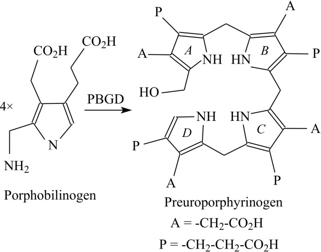
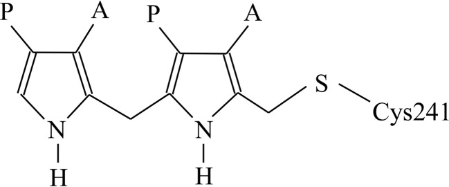
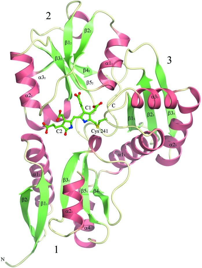
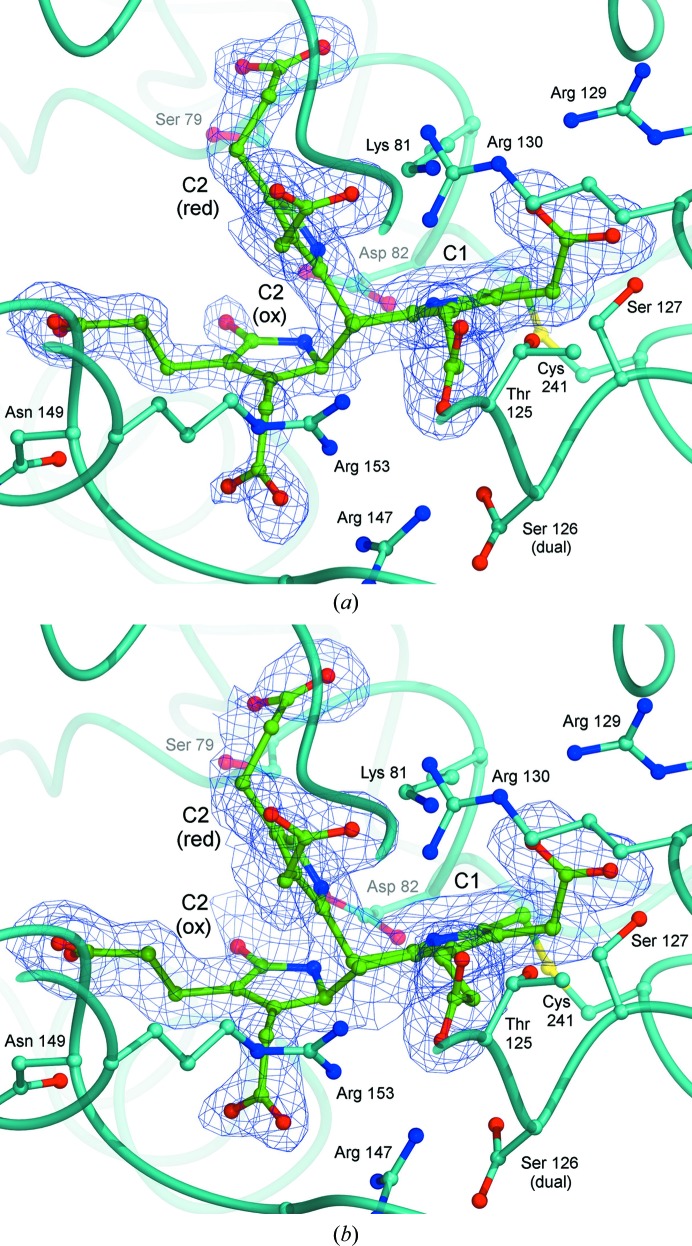
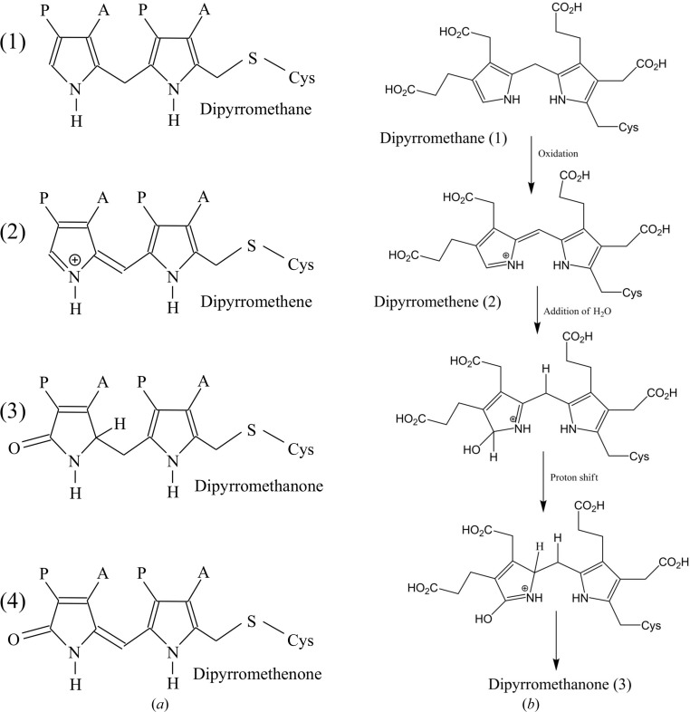
The enzyme porphobilinogen deaminase (PBGD; hydroxymethylbilane synthase; EC 2.5.1.61) catalyses an early step of the tetrapyrrole-biosynthesis pathway in which four molecules of the monopyrrole porphobilinogen are condensed to form a linear tetrapyrrole. The enzyme possesses a dipyrromethane cofactor, which is covalently linked by a thioether bridge to an invariant cysteine residue (Cys241 in the Bacillus megaterium enzyme). The cofactor is extended during the reaction by the sequential addition of the four substrate molecules, which are released as a linear tetrapyrrole product. Expression in Escherichia coli of a His-tagged form of B. megaterium PBGD has permitted the X-ray analysis of the enzyme from this species at high resolution, showing that the cofactor becomes progressively oxidized to the dipyrromethene and dipyrromethanone forms. In previously solved PBGD structures, the oxidized cofactor is in the dipyromethenone form, in which both pyrrole rings are approximately coplanar. In contrast, the oxidized cofactor in the B. megaterium enzyme appears to be in the dipyrromethanone form, in which the C atom at the bridging α-position of the outer pyrrole ring is very clearly in a tetrahedral configuration. It is suggested that the pink colour of the freshly purified protein is owing to the presence of the dipyrromethene form of the cofactor which, in the structure reported here, adopts the same conformation as the fully reduced dipyrromethane form.

Keywords: dipyrromethane cofactor; porphobilinogen deaminase; tetrapyrrole biosynthesis.


Figures

Figure 1
Figure 1
The reaction catalysed by porphobilinogen deaminase. Four molecules of the pyrrole porphobilinogen are condensed to form the linear tetrapyrrole preuroporphyrinogen (hydroxymethylbilane). The acetic acid and propionic acid side chains of each pyrrole are abbreviated A and P, respectively, and the four rings of the tetrapyrrole product are indicated in italics as A, B, C and D.
Figure 2
Figure 2
The dipyrromethane cofactor of porphobilinogen deaminase is covalently attached to the enzyme by a thioether bond to a cysteine residue. Four substrate pyrroles are added linearly to the cofactor; finally, hydrolysis of the linkage between the substrate and the cofactor releases the tetrapyrrole product hydroxymethylbilane.
Figure 3
Figure 3
The tertiary structure of B. megaterium PBGD at 1.46 Å resolution showing the oxidized form of the dipyrromethane cofactor covalently attached to Cys241. The domains of the enzyme are numbered 1–3 and the secondary-structure elements are labelled according to the nomenclature of Louie et al. (1992 ▶).
Figure 4
Figure 4
Sequence alignment and secondary structure of B. megaterium PBGD. An alignment of B. megaterium PBGD with the enzyme from another prokaryote (E. coli) along with the plant (A. thaliana) and human enzymes. The secondary-structure elements are labelled using the notation of Louie et al. (1992 ▶) and the amino-acid residues are colour-coded as follows: cyan, basic; red, acidic; green, neutral polar; pink, bulky hydrophobic; white, Gly, Ala and Pro; yellow, Cys.
Figure 5
Figure 5
Superposition of B. megaterium PBGD with the Arabidopsis, E. coli and human enzymes. (a) The overall least-squares superposition of B. megaterium PBGD (cyan) with the E. coli enzyme (yellow) as well as the Arabidopsis and human enzymes (green and pink, respectively). A superposition of the four enzymes based on domain 1 alone is shown in (b), which emphasizes the different concerted shifts of domains 2 and 3 relative to domain 1 in the enzyme from each species.
Figure 6
Figure 6
Electron density showing the dual conformations of the cofactor. The local fold of the protein is indicated as a cyan tube and the side chains adjacent to the cofactor are shown in the same colour as the enzyme, while the cofactor itself is coloured green. The map obtained for the 40-­day-old protein is shown in (a), with that for the 50-day-old protein shown in (b); both maps are contoured at 0.7 r.m.s.. The C2 ring clearly adopts two positions depending on its oxidation state, which are shown as C2(ox) and C2(red). In the oxidized conformation, the C2 ring possesses a carbonyl O atom substituted at the α-position; accordingly, the proportion of cofactor adopting this conformer increases with time.
Figure 7
Figure 7
Oxidation states of the dipyrromethane cofactor. Different possible oxidation states of the dipyrromethane cofactor are shown in (a) along with a proposed mechanism for oxidation of the dipyrromethane to dipyrromethene and subsequently dipyrromethanone (b).

References

    1. Awan, S. J., Siligardi, G., Shoolingin-Jordan, P. M. & Warren, M. J. (1997). Biochemistry, 36, 9273–9282. - PubMed
    1. Azim, N., Deery, E., Warren, M. J., Erskine, P., Cooper, J. B., Wood, S. P. & Akhtar, M. (2013). Acta Cryst. F69, 906–908. - PMC - PubMed
    1. Battersby, A. R., Fookes, C. J. R., Hart, G., Matcham, G. W. J. & Pandey, P. S. (1983). J. Chem. Soc. Perkin Trans. 1, 1983, 3041–3047.
    1. Bunk, B., Schulz, A., Stammen, S., Münch, R., Warren, M. J., Rohde, M., Jahn, D. & Biedendieck, R. (2010). Bioeng. Bugs, 1, 85–91. - PMC - PubMed
    1. Chen, V. B., Arendall, W. B., Headd, J. J., Keedy, D. A., Immormino, R. M., Kapral, G. J., Murray, L. W., Richardson, J. S. & Richardson, D. C. (2010). Acta Cryst. D66, 12–21. - PMC - PubMed

Publication types

MeSH terms