. 2014 Mar 5;9(3):e90829.
doi: 10.1371/journal.pone.0090829. eCollection 2014.

Preferential macrophage recruitment and polarization in LPS-induced animal model for COPD: noninvasive tracking using MRI

Affiliations

Preferential macrophage recruitment and polarization in LPS-induced animal model for COPD: noninvasive tracking using MRI

Achraf Al Faraj et al. PLoS One. .

Abstract

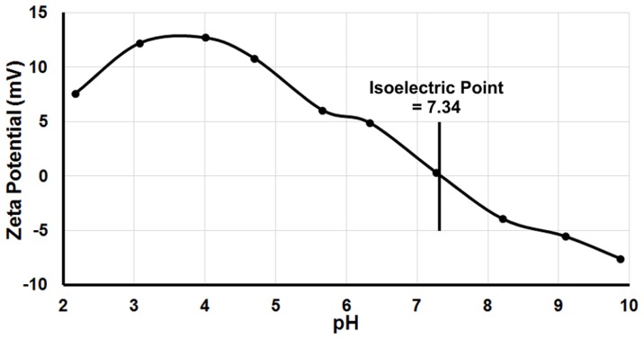
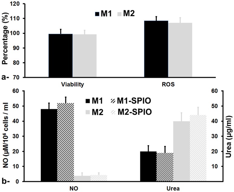
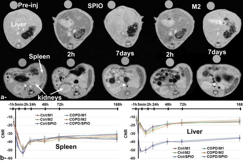
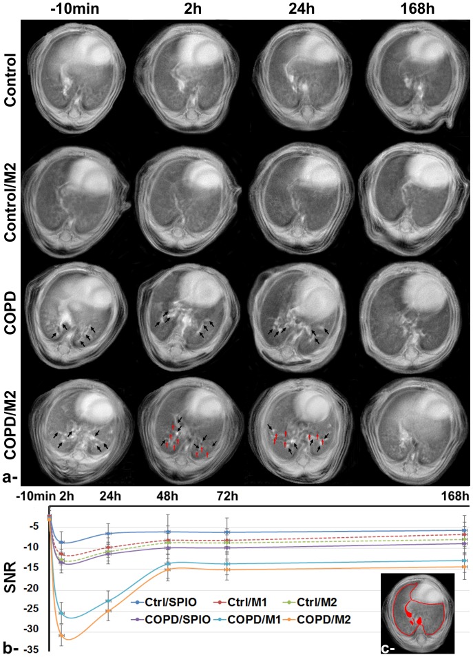
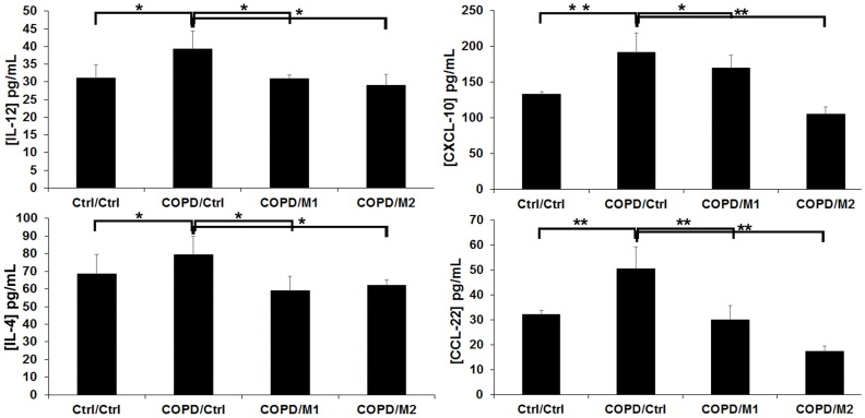
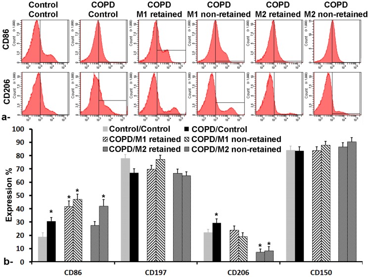
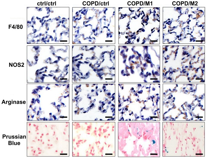
Noninvasive imaging of macrophages activity has raised increasing interest for diagnosis of chronic obstructive respiratory diseases (COPD), which make them attractive vehicles to deliver contrast agents for diagnostic or drugs for therapeutic purposes. This study was designed to monitor and evaluate the migration of differently polarized M1 and M2 iron labeled macrophage subsets to the lung of a LPS-induced COPD animal model and to assess their polarization state once they have reached the inflammatory sites in the lung after intravenous injection. Ex vivo polarized bone marrow derived M1 or M2 macrophages were first efficiently and safely labeled with amine-modified PEGylated dextran-coated SPIO nanoparticles and without altering their polarization profile. Their biodistribution in abdominal organs and their homing to the site of inflammation in the lung was tracked for the first time using a free-breathing non-invasive MR imaging protocol on a 4.7T magnet after their intravenous administration. This imaging protocol was optimized to allow both detection of iron labeled macrophages and visualization of inflammation in the lung. M1 and M2 macrophages were successfully detected in the lung starting from 2 hours post injection with no variation in their migration profile. Quantification of cytokines release, analysis of surface membrane expression using flow cytometry and immunohistochemistry investigations confirmed the successful recruitment of injected iron labeled macrophages in the lung of COPD mice and revealed that even with a continuum switch in the polarization profile of M1 and M2 macrophages during the time course of inflammation a balanced number of macrophage subsets predominate.


Conflict of interest statement

Competing Interests: The authors have declared that no competing interests exist.

Figures

Figure 1
Figure 1. Zeta potential of amine-modified PEGylated dextran-coated iron oxide nanoparticles assessed in ultrapure water at 25°C.
Figure 2
Figure 2. Relative percentage of viability and reactive oxygen species generation of M1 and M2 SPIO labeled macrophages compared to unlabeled macrophages subsets after the overnight chase period (a).
Nitric oxide (NO) release as marker of iNOS activity (left) in M1 macrophages and Arginine-derived urea production (right) as marker of Arginase1 activity in M2 macrophages (b). Error bars are standard deviation of triplicates.
Figure 3
Figure 3. MR images acquired using susceptibility-weighted gradient echo sequence showing the liver (upper row) and the spleen and kidneys (lower row) pre-injection (−1 h) and at 2 hours and 7 days post-injection of either free SPIO or SPIO labeled M2 macrophages in control mice groups (a).
Contrast-to-noise (CNR) variation during the 7 days follow-up study for the spleen (left side) and the liver (right side) before and after intravenous injection of either free SPIO, SPIO labeled M1 or M2 macrophages in control and COPD animal groups (b). Error bars are standard deviation of triplicates.
Figure 4
Figure 4. MR Images acquired using ultra-short echo time (UTE) sequence of control and LPS-induced COPD lungs, with or without injection of M2 macrophages (a).
From top to bottom: Control, Control/M2, COPD, COPD/M2 groups imaged at (from left to right) −10 min, 2 h, 24 h and 168 h post M2 macrophages injection. Black arrows highlight the inflammatory regions in COPD groups and red arrows highlight the presence of void signal dots related to higher macrophage infiltrations in the inflammatory lungs. Signal-to-noise (SNR) attenuation of lung parenchyma, during the 7 days follow-up study, measured before and after intravenous injection of either free SPIO, SPIO labeled M1 or M2 macrophages in control and COPD mice (b). Error bars are standard deviation of triplicates. Representative regions of interest (ROI) which were drawn around apparent vascular structures (filled in red) and subtracted from the map to retain lung parenchyma (c).
Figure 5
Figure 5. Interleukins (IL-12, IL-4) and Chemokines (CCL-22 and CXCL-10) levels quantified by ELISA assay obtained from BAL samples of ctrl/ctrl, COPD/ctrl, COPD/M1 and COPD/M2 animal groups at 2 h post macrophage injection.
Data expressed as mean ± SD, n = 5 per group. *p<0.05; **p<0.01.
Figure 6
Figure 6. Flow cytometry analysis of alveolar macrophages in control/control, COPD/Control (48 h post LPS intrapulmonary exposition), COPD/M1 and COPD/M2 (2 h post-injection of iron-labeled macrophages) groups for both retained (i.e., iron loaded) and retained fractions.
a- representative histogram of CD86 expression (higher row) and CD206 (lower row). b- Surface membrane receptor expression percentage of CD86, CD197, CD206 and CD150. Error bars are standard deviation of triplicates. *p<0.05.
Figure 7
Figure 7. Examples of Immunohistochemical (IHC) staining of lungs sections of ctrl/ctrl, COPD/ctrl, COPD/M1 and COPD/M2 groups at 2 hours post-injection of macrophages using: F4/80 (first row) as universal marker for macrophages, iNOS (second row) and Arginase1 (third row) as marker for M1 and M2 macrophages respectively and of Prussian blue iron staining (fourth row) in adjacent sections revealing the presence of iron oxide nanoparticles (blue dots) in macrophages injected groups.
Scale bar: 100 µm.

References

    1. Global strategy for the diagnosis, management and prevention of chronic obstructive pulmonary disease. Global Initiative for Obstructive Lung Disease. Available: http://www.goldcopd.com. Accessed 2013 Feb 20: GOLD 2013.
    1. Barnes P (2004) Alveolar Macrophages as Orchestrators of COPD. COPD: Journal of Chronic Obstructive Pulmonary Disease 1: 59–70. - PubMed
    1. Herold S, Mayer K, Lohmeyer J (2011) Acute lung injury: how macrophages orchestrate resolution of inflammation and tissue repair. Front Immunol 2: 65. - PMC - PubMed
    1. Biswas SK, Chittezhath M, Shalova IN, Lim JY (2012) Macrophage polarization and plasticity in health and disease. Immunol Res 53: 11–24. - PubMed
    1. Mosser DM, Edwards JP (2008) Exploring the full spectrum of macrophage activation. Nat Rev Immunol 8: 958–969. - PMC - PubMed

Publication types

MeSH terms