Review
. 2014 Jun:30:2-13.
doi: 10.1016/j.semcdb.2014.02.012. Epub 2014 Mar 2.

The regulation of spermatogenesis by androgens

Affiliations
Review

The regulation of spermatogenesis by androgens

Lee B Smith et al. Semin Cell Dev Biol. 2014 Jun.

Abstract

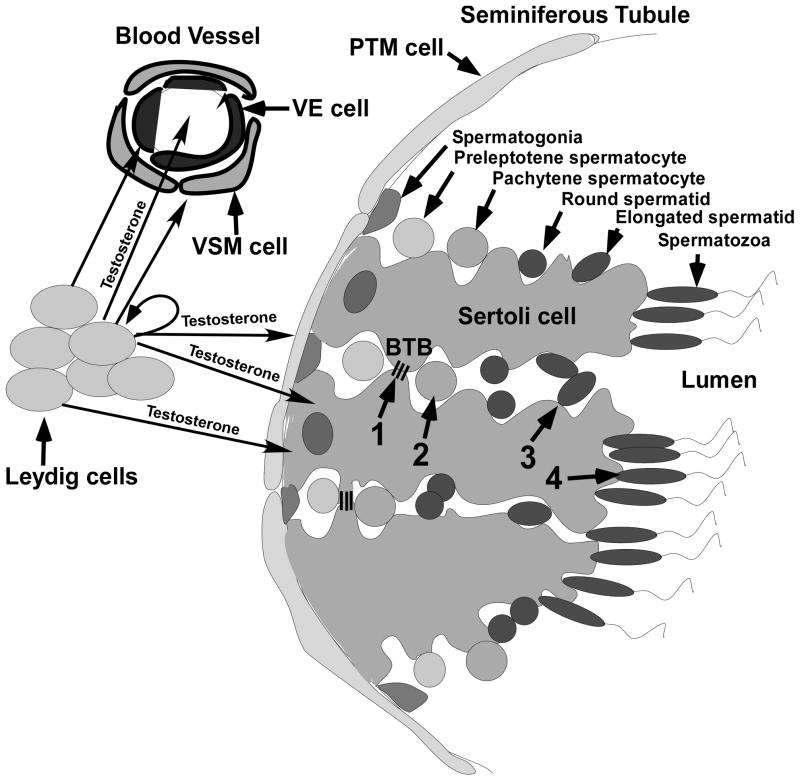
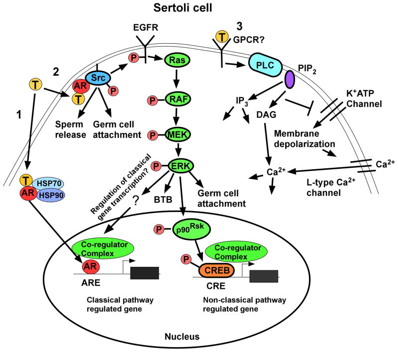
Testosterone is essential for maintaining spermatogenesis and male fertility. However, the molecular mechanisms by which testosterone acts have not begun to be revealed until recently. With the advances obtained from the use of transgenic mice lacking or overexpressing the androgen receptor, the cell specific targets of testosterone action as well as the genes and signaling pathways that are regulated by testosterone are being identified. In this review, the critical steps of spermatogenesis that are regulated by testosterone are discussed as well as the intracellular signaling pathways by which testosterone acts. We also review the functional information that has been obtained from the knock out of the androgen receptor from specific cell types in the testis and the genes found to be regulated after altering testosterone levels or androgen receptor expression.

Keywords: Blood testis barrier; Fertility; Meiosis; Sertoli cell; Testis; Testosterone.


Figures

Figure 1
Figure 1
The anatomy of the testis and the process of spermatogenesis. Leydig cells and blood vessels that are lined by vascular endothelial (VE) and vascular smooth muscle (VSM) cells are localized to the interstitial space between seminiferous tubules. Peritubular myoid (PTM) cells line the outside of the seminiferous tubule. Sertoli cells extend from the PTM cells to the lumen and surround the developing germ cells. The process of spermatogenesis is outlined at the top of the figure where germ cell development is shown progressing toward the lumen from spermatogonia on the basement membrane to the production of spermatozoa. Testosterone produced by the Leydig cells diffuses into the VE and VSM cells as well as into the blood vessels. Testosterone also diffuses from the Leydig cells into PTM, Sertoli cells and Leydig cells. The four critical processes regulated by testosterone (1–4 in the middle of the figure) are indicated: 1) the maintenance of the BTB (represented by 3 lines extending between 2 Sertoli cells), 2) completion of meiosis by spermatocytes, 3) adherence of elongated spermatids to Sertoli cells and 4) the release of mature spermatozoa.
Figure 2
Figure 2
Testosterone signaling pathways in Sertoli cells. Left (Pathway 1): The classical testosterone signaling pathway: Testosterone diffuses through the plasma membrane and binds with the AR that then undergoes an alteration in conformation allowing it to be released from heat shock proteins (HSP70, HSP90) in the cytoplasm. AR then translocates to the nucleus where it binds to specific DNA sequences called androgen response elements (AREs). AR binding to an ARE allows the recruitment of co-regulators (co-activator or co-repressor proteins) that alter the expression of genes and eventually cellular function. Middle (Pathway 2): The non-classical kinase activation pathway: Testosterone interacts with AR that is then able to bind with and activate Src, which is required for sperm release and germ cell attachment. Src also causes the activation of the EGF receptor that then activates the MAP kinase cascade most likely through Ras resulting in the sequential phosphorylation and activation of RAF and MEK and then ERK, which regulates BTB integrity and germ cell attachment. ERK may also regulate classical pathway-mediated gene expression through pathways that are not yet known for Sertoli cells. ERK also can facilitate non-classical pathway mediated gene expression. For example, ERK activates p90RSK kinase, which is known to phosphorylate CREB on serine 133. As a result, CREB-regulated genes can be induced by testosterone. Right (Pathway 3): The non-classical Ca2+ influx pathway: Testosterone interacts with a receptor in the plasma membrane that has characteristics of a Gq coupled G-protein coupled receptor (GPCR). Phospholipase C (PLC) is activated to cleave PIP2 into IP3 and DAG. Decreased PIP2 concentrations inhibit K+ATP channels causing membrane depolarization and Ca2+ entry via L-type Ca2+ channels.

References

    1. Chang C, Lee SO, Wang RS, Yeh S, Chang TM. Androgen receptor (AR) physiological roles in male and female reproductive systems: lessons learned from AR-knockout mice lacking AR in selective cells. Biol Reprod. 2013;89:21. - PMC - PubMed
    1. Walker WH. Non-classical actions of testosterone and spermatogenesis. Philos Trans R Soc Lond B Biol Sci. 2010;365:1557–69. - PMC - PubMed
    1. Walker WH. Testosterone signaling and the regulation of spermatogenesis. Spermatogenesis. 2011;1:116–120. - PMC - PubMed
    1. Verhoeven G, Willems A, Denolet E, Swinnen JV, De Gendt K. Androgens and spermatogenesis: lessons from transgenic mouse models. Philos Trans R Soc Lond B Biol Sci. 2010;365:1537–56. - PMC - PubMed
    1. Alves MG, Rato L, Carvalho RA, Moreira PI, Socorro S, Oliveira PF. Hormonal control of Sertoli cell metabolism regulates spermatogenesis. Cell Mol Life Sci. 2013;70:777–93. - PMC - PubMed

Publication types

LinkOut - more resources