. 2014 Mar 5;9(3):e90429.
doi: 10.1371/journal.pone.0090429. eCollection 2014.

Effects of blood contamination and the rostro-caudal gradient on the human cerebrospinal fluid proteome

Affiliations

Effects of blood contamination and the rostro-caudal gradient on the human cerebrospinal fluid proteome

Elise Aasebø et al. PLoS One. .

Abstract

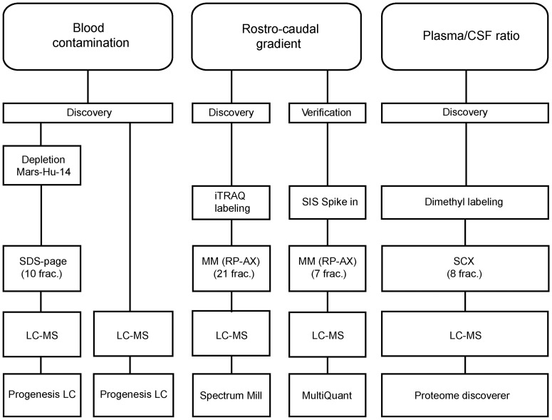
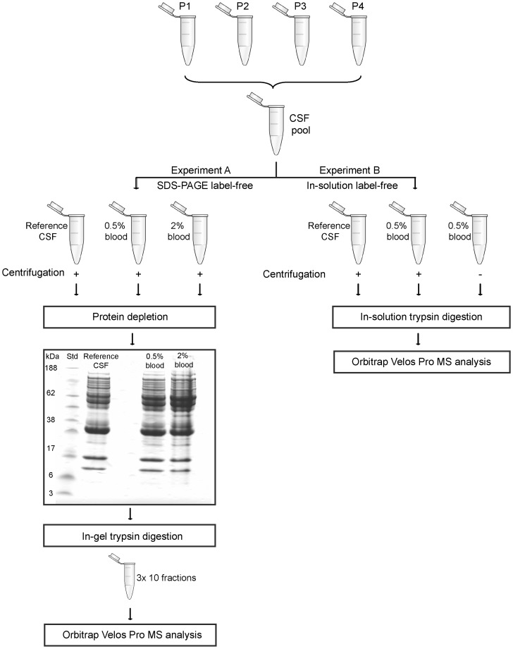
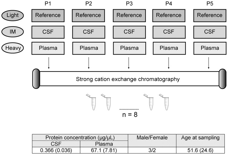
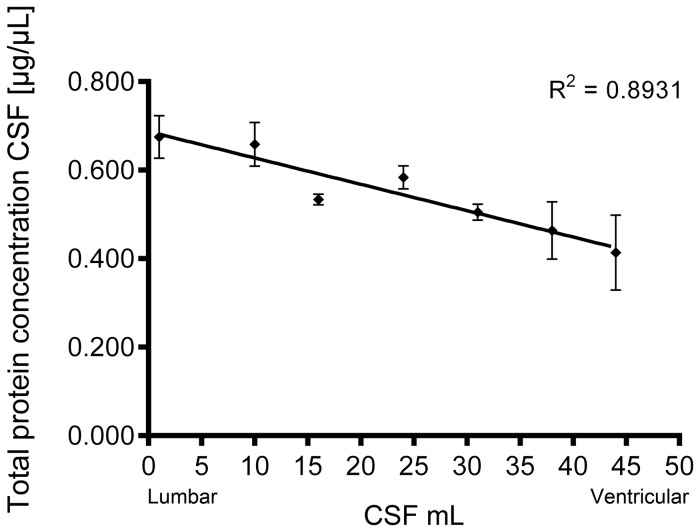
Over the last years there has been an increased focus on the importance of knowing the effect of pre-analytical influence on the proteomes under study, particularly in the field of biomarker discovery. We present three proteomics studies examining the effect of blood contamination and the rostro-caudal gradient (RCG) on the cerebrospinal fluid (CSF) proteome, in addition to plasma/CSF protein ratios. The studies showed that the central nervous system (CNS) derived proteins appeared to be unaffected by the RCG, while the plasma-derived proteins showed an increase in concentration towards the lumbar area. This implies that the concentration of the plasma-derived proteins in CSF will vary depending on the volume of CSF that is collected. In the CSF samples spiked with blood, 262 of 814 quantified proteins showed an abundance increase of more than 1.5 fold, while 403 proteins had a fold change of less than 1.2 and appeared to be unaffected by blood contamination. Proteins with a high plasma/CSF ratio appeared to give the largest effect on the CSF proteome upon blood contamination. The results give important background information on how factors like blood contamination, RCG and blood-CNS-barrier influences the CSF proteome. This information is particularly important in the field of biomarker discovery, but also for routine clinical measurements. The data from the blood contamination and RCG discovery studies have been deposited to the ProteomeXchange with identifier PXD000401.


Conflict of interest statement

Competing Interests: The authors have declared that no competing interests exist.

Figures

Figure 1
Figure 1. Overview of the conducted studies.
Blood contamination: in experiment A protein depleted CSF was separated by SDS-PAGE, in experiment B crude CSF was in-solution digested. Progenesis LC-MS was used for data analysis. In the rostro-caudal gradient study, we used iTRAQ-labeling with mixed mode reversed phase-anion chromatography (MM (RP-AX)) fractionation. The Spectrum Mill software was used for data analysis. For verification we used stable isotope dilution (SID) multiple reaction monitoring (MRM) to monitor 70 peptides. MM (RP-AX) chromatography was used for fractionation and the MultiQuant software was used for data analysis. In the plasma/CSF ratio study equal amount of corresponding CSF and plasma from five patients were compared using dimethyl labeling. Samples were fractionated using strong cation exchange (SCX) chromatography. Proteome discoverer was used for data analysis. For all discovery experiments the samples were analyzed on an Orbitrap Velos Pro and for SID-MRM the samples were analyzed on a Q-trap 5500. SIS = Stable Isotope Standards
Figure 2
Figure 2. Flow chart of the blood contamination studies.
Cerebrospinal fluid (CSF) from four patients (P1-P4) were pooled and divided into aliquots which were spiked with different amounts of blood. In experiment A, a reference sample was compared to CSF spiked to achieve 0.5% and 2% blood. The CSF samples were centrifuged, protein depleted, and separated by SDS-PAGE into ten fractions per lane. In experiment B, we examined the effect of centrifugation. A reference sample was compared to two CSF samples spiked to achieve 0.5% blood, where one of the blood contaminated samples was not centrifuged. The samples were trypsin digested in-solution. All samples were analyzed on an OrbiTrap Velos Pro.
Figure 3
Figure 3. Flow chart of the rostro-caudal gradient study.
In the rostro-caudal gradient (RCG) study, we examined the seven following points of the RCG from a PSP patient: 1-2nd, 10-11th, 16-17th, 24-25th, 31-32nd, 38-39th and 44-45th mL CSF, referred to as RCG point 1-7, respectively. Twelve samples were digested and iTRAQ labeled (114-117). A reference, (labeled with iTRAQ reagent 114) containing the same amount of each RCG point, was included in each experiment. The RCG points 1 and 7 were included twice. After digestion and iTRAQ-labeling, samples were combined as follows: Exp. 1 (reference, 44-45th mL, 24-25th mL and 1-2nd mL), Exp. 2 (reference, 1-2nd mL, 38-39th mL and 16-17th mL), and Exp. 3 (reference, 10-11th mL, 44-45th mL and the 31-32nd mL). The three experiments were fractionated by mixed mode reversed phase-anion chromatography (MM (RP-AX)) and analyzed on an Orbitrap Velos Pro. The protein abundances were averaged for each protein in the duplicate samples.
Figure 4
Figure 4. Flow chart of the plasma/CSF study.
We compared the cerebrospinal fluid (CSF) and plasma protein ratio of five patients (P1-P5) using dimethyl labeling. The reference sample was a mix of equal total amount of CSF and plasma, and was labeled by light reagents. The five CSF samples were labeled by intermediate (IM) reagents, and the plasma samples were labeled by the heavy reagents. The light, IM and heavy labeled samples were combined and fractionated into eight fractions by strong cation exchange chromatography and analyzed on an Orbitrap Velos Pro. The average (and standard deviation) protein concentration of CSF and plasma, age at sampling and ratio male/female of the five patients are presented in the figure.
Figure 5
Figure 5. Protein concentration measurement of along the rostro-caudal gradient.
Protein concentration of the cerebrospinal fluid (CSF)-derived proteins from the seven points along the rostro-caudal gradient (RCG). The CSF was measured in triplicates with Qubit, and error bars of the standard deviation are included. R squared value 0.8931.

References

    1. Simonsen AH, Bahl JM, Danborg PB, Lindstrom V, Larsen SO, et al. (2013) Pre-analytical factors influencing the stability of cerebrospinal fluid proteins. J Neurosci Methods 215: 234–240. - PubMed
    1. Berven FS, Kroksveen AC, Berle M, Rajalahti T, Flikka K, et al. (2007) Pre-analytical influence on the low molecular weight cerebrospinal fluid proteome. Proteomics Clinical Applications 1: 699–711. - PubMed
    1. Jimenez CR, Koel-Simmelink M, Pham TV, van der Voort L, Teunissen CE (2007) Endogeneous peptide profiling of cerebrospinal fluid by MALDI-TOF mass spectrometry: Optimization of magnetic bead-based peptide capture and analysis of preanalytical variables. Proteomics Clin Appl 1: 1385–1392. - PubMed
    1. Teunissen CE, Petzold A, Bennett JL, Berven FS, Brundin L, et al. (2009) A consensus protocol for the standardization of cerebrospinal fluid collection and biobanking. Neurology 73: 1914–1922. - PMC - PubMed
    1. Petzold A, Sharpe LT, Keir G (2006) Spectrophotometry for cerebrospinal fluid pigment analysis. Neurocrit Care 4: 153–162. - PubMed

Publication types

MeSH terms