. 2014 Mar 5;9(3):e90671.
doi: 10.1371/journal.pone.0090671. eCollection 2014.

Differential neuroprotective effects of 5'-deoxy-5'-methylthioadenosine

Affiliations

Differential neuroprotective effects of 5'-deoxy-5'-methylthioadenosine

Beatriz Moreno et al. PLoS One. .

Abstract

Background: 5'-deoxy-5'-methylthioadenosine (MTA) is an endogenous compound produced through the metabolism of polyamines. The therapeutic potential of MTA has been assayed mainly in liver diseases and, more recently, in animal models of multiple sclerosis. The aim of this study was to determine the neuroprotective effect of this molecule in vitro and to assess whether MTA can cross the blood brain barrier (BBB) in order to also analyze its potential neuroprotective efficacy in vivo.

Methods: Neuroprotection was assessed in vitro using models of excitotoxicity in primary neurons, mixed astrocyte-neuron and primary oligodendrocyte cultures. The capacity of MTA to cross the BBB was measured in an artificial membrane assay and using an in vitro cell model. Finally, in vivo tests were performed in models of hypoxic brain damage, Parkinson's disease and epilepsy.

Results: MTA displays a wide array of neuroprotective activities against different insults in vitro. While the data from the two complementary approaches adopted indicate that MTA is likely to cross the BBB, the in vivo data showed that MTA may provide therapeutic benefits in specific circumstances. Whereas MTA reduced the neuronal cell death in pilocarpine-induced status epilepticus and the size of the lesion in global but not focal ischemic brain damage, it was ineffective in preserving dopaminergic neurons of the substantia nigra in the 1-methyl-4-phenyl-1,2,3,6-tetrahydro-pyridine (MPTP)-mice model. However, in this model of Parkinson's disease the combined administration of MTA and an A2A adenosine receptor antagonist did produce significant neuroprotection in this brain region.

Conclusion: MTA may potentially offer therapeutic neuroprotection.


Conflict of interest statement

Competing Interests: The authors have declared that no competing interests exist.

Figures

Figure 1
Figure 1. Effect of MTA under NMDA excitotoxicity.
Effect of MTA co-treatment (a) or MTA pre-treatment (b) on NMDA-induced caspase 3 activation in rat pure primary neuronal cultures. Effect of MTA co-treatment (c) or MTA pre-treatment (d) on NMDA-induced caspase 3 activation in rat mixed astrocyte-neuron cultures. Caspase 3 activity (units of fluorescence per milligram of protein per hour) was determined in cells treated with 300 µM NMDA, in the presence or absence of MTA (panels a and b: 100, 250 and 500 µM; panels c and d: 250 µM) or 10 µM MK-801 (MK; NMDA receptor antagonist). The results are expressed as the mean ± SEM of at least four independent experiments performed in triplicate: *p<0.05, **p<0.01***p <0.001 compared with cells treated with NMDA; # p<0.05, ## p<0.01, ### p<0.001 compared with vehicle cells. One-way analysis of variance (ANOVA) and Bonferroni's t-test for multiple comparisons.
Figure 2
Figure 2. Effect of MTA under OGD and AMPA excitotoxicity.
Panels a and b: the effect of APV (100 µM) or MTA in OGD conditions in rat mixed cultures of neurons and astrocytes. Cell death was expressed as the LDH activity at the beginning of any treatment: *p<0.05 compared to IAA-treated cells; # p<0.05 compared to control; ** p<0.01 compared to IAA-treated cells; ## p<0.01 compared to control. c) primary oligodendrocytes derived from rat optic nerve subjected to AMPA excitotoxicity (10 µM and 100 µM) in which cell death was measured with calcein-AM, as indicated in Methods. *p<0.05 compared with cells treated with AMPA (Unpaired t-test). Values represent the average ± SEM and were obtained from at least three independent experiments performed in duplicates.
Figure 3
Figure 3. Effect of MTA on brain ischemia.
(a) Representative TTC stained sections of vehicle and MTA treated animals (30 mg/kg/twice daily, i.p.) 3 days after the induction of transient focal ischemia. Histogram (right) showing the infarct volume calculated from TTC stained slices in vehicle- and MTA-treated rats (n = 5 in each group). b) Representative microphotographs of Fluoro Jade C staining after rat transient forebrain ischemia (n = 5 in the vehicle group; n = 6 in the MTA-treated group). MTA (30 mg/kg) was administered 30 min after triggering ischemia. Quantification of Fluoro Jade positive cells per mm length of CA1 pyramidal layer (right). The data represents the mean ± SEM: **p<0.01 compared to the vehicle (Student t-test). Scale bar 100 µm.
Figure 4
Figure 4. MTA effects in chronic pilocarpine-induced status epilepticus (SE).
a–f: Representative images of Neu N immunoreactivity in the hippocampus are shown 3 days after sham treatment (a) or pilocarpine-induced SE (b, c), or 30 days after SE (d–f). MTA (30 mg/kg) was administered pre-SE (d) or post-SE (c,f) induction. Cell loss is already apparent by 3 days (arrows in b) and is marked by 30 days (e) after SE, but it appears attenuated in MTA-treated animals at both timepoints (c,d,f). g–h) Bar plots show quantification of NeuN-positive cells in CA3(g) or CA1(h) 30 days after SE in animals pre-treated with MTA. The data represent the mean ± SEM. *p<0.05; **p<0.001; ANOVA with Tukey HSD post –hoc test.
Figure 5
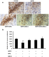
Figure 5. Effect of MTA and A2AR antagonists in an acute mouse model of PD.
The effect of MTA (30 mg/kg), MSX-3 (9 mg/kg) or both in combination on the survival of dopaminergic neurons (TH+ cells) within the substantia nigra of the brain of mice treated with MPTP. a) Representative images of the neurodegeneration of dopaminergic cells in the different groups. b) Stereological counts of TH+ neurons in control and drug-treated animals (n = 6 per group). The data represent the mean±SEM: *p<0.05 comparing with the MPTP group; # p<0.05 comparing with the control group (One-way ANOVA, LSD test as a post-hoc). Scale bar: 50 µm.

References

    1. Williams-Ashman HG, Seidenfeld J, Galletti P (1982) Trends in the biochemical pharmacology of 5′-deoxy-5′-methylthioadenosine. Biochem Pharmacol 31(3): 277–88. - PubMed
    1. Avila MA, García-Trevijano ER, Lu SC, Corrales FJ, Mato JM (2004) Methylthioadenosine. Int J Biochem Cell Biol 36(11): 2125–30. - PubMed
    1. Latasa MU, Gil-Puig C, Fernández-Barrena MG, Rodríguez-Ortigosa CM, Banales JM, et al. (2010) Oral Methylthioadenosine Administration Attenuates Fibrosis and Chronic Liver Disease Progression in Mdr22/2 Mice. PLoS ONE 5(12): e15690 doi:10.1371/journal.pone.0015690 - DOI - PMC - PubMed
    1. Moreno B, Hevia H, Santamaria M, Sepulcre J, Muñoz J, et al. (2006) Methylthioadenosine reverses brain autoimmune disease. Ann Neurol 60(3): 323–34. - PubMed
    1. Moreno B, Fernandez-Diez B, Di Penta A, Villoslada P (2010) Preclinical studies of methylthioadenosine for the treatment of multiple sclerosis. Mult Scler 16(9): 1102–8. - PubMed

Publication types

MeSH terms