. 2014 Mar 5;34(10):3607-21.
doi: 10.1523/JNEUROSCI.3388-13.2014.

Acute suppressive and long-term phase modulation actions of orexin on the mammalian circadian clock

Affiliations

Acute suppressive and long-term phase modulation actions of orexin on the mammalian circadian clock

Mino D C Belle et al. J Neurosci. .

Abstract

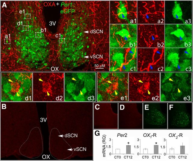
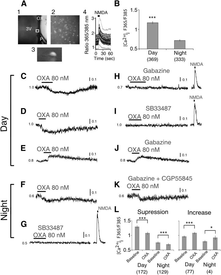
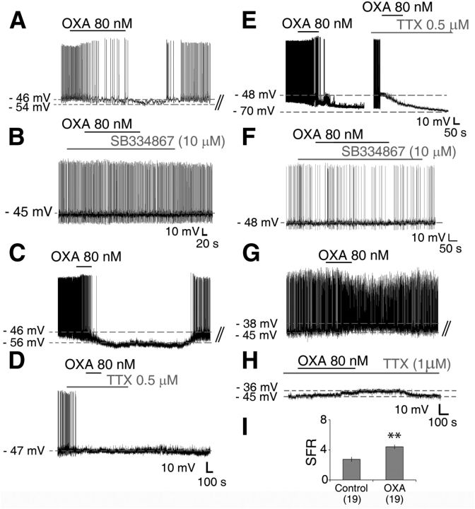
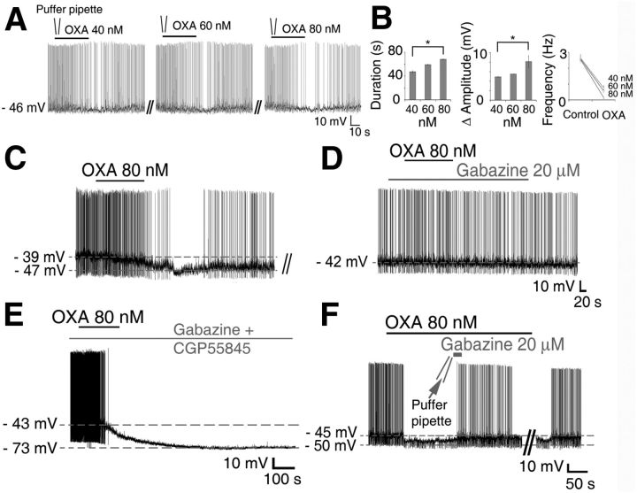
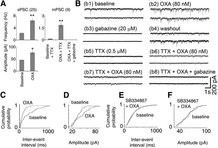
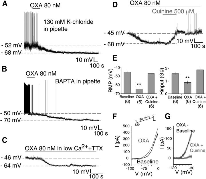
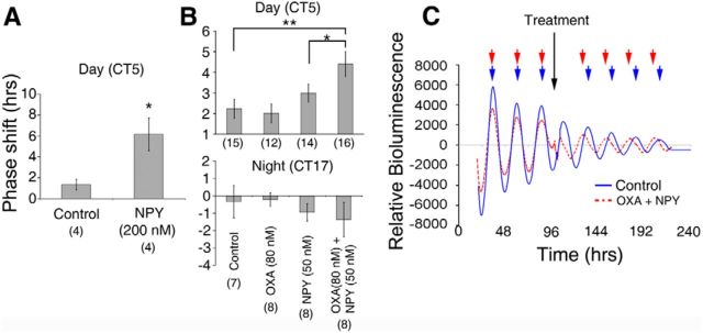
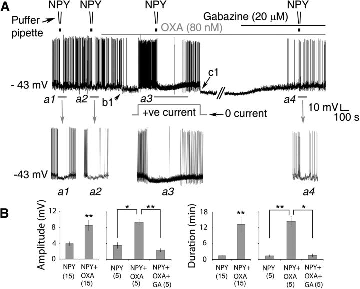
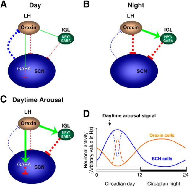
Circadian and homeostatic neural circuits organize the temporal architecture of physiology and behavior, but knowledge of their interactions is imperfect. For example, neurons containing the neuropeptide orexin homeostatically control arousal and appetitive states, while neurons in the suprachiasmatic nuclei (SCN) function as the brain's master circadian clock. The SCN regulates orexin neurons so that they are much more active during the circadian night than the circadian day, but it is unclear whether the orexin neurons reciprocally regulate the SCN clock. Here we show both orexinergic innervation and expression of genes encoding orexin receptors (OX1 and OX2) in the mouse SCN, with OX1 being upregulated at dusk. Remarkably, we find through in vitro physiological recordings that orexin predominantly suppresses mouse SCN Period1 (Per1)-EGFP-expressing clock cells. The mechanisms underpinning these suppressions vary across the circadian cycle, from presynaptic modulation of inhibitory GABAergic signaling during the day to directly activating leak K(+) currents at night. Orexin also augments the SCN clock-resetting effects of neuropeptide Y (NPY), another neurochemical correlate of arousal, and potentiates NPY's inhibition of SCN Per1-EGFP cells. These results build on emerging literature that challenge the widely held view that orexin signaling is exclusively excitatory and suggest new mechanisms for avoiding conflicts between circadian clock signals and homeostatic cues in the brain.

Keywords: GABA; arousal; circadian; electrophysiology; orexin A; suprachiasmatic.


Figures

Figure 1.
Figure 1.
Neurons of the SCN are surrounded by OXA-containing axons and encode for orexin receptors. A, Collapsed stacks of confocal images taken from the middle of the rostrocaudal axis of the SCN of Per1-EGFP mice, and immunostained with anti-OXA. A, Shows OXA-ir fiber distribution (red) in the SCN with Per1-EGFP neurons (green) delineating the dorsal (dSCN) and ventral (vSCN) anatomical structure of the SCN. a1e3, Montage of merged and collapsed confocal image stacks of Per1-EGFP neurons (green) and OXA-ir (red) at 63× magnification (taken from regions in A marked by white dotted boxes). Insets, a1c1 show orexinergic fibers (yellow) on Per1-EGFP cells (blue arrows), with a2c2 showing separate OXA-ir axons (red) and a3c3 Per1-EGFP neurons. Insets, d1 and e1 show examples of OXA-ir axonal boutons converging on two cells that did not contain EGFP (yellow arrows). d2 and e2 show the same two cells with the green channel disabled, and d3 and e3 confirm the absence of EGFP in these two cells close to OXA-ir varicotic fibers. Visual confirmation of OXA-ir varicotic fibers and terminals on Per1-EGFP cells was performed on 3D rotated projections of optical confocal image slices before image stack collapsing (see Materials and Methods). B, Confocal image showing the absence of OXA-ir staining in the SCN and adjacent hypothalamic areas of a prepro-orexin knock-out mouse, while C and D show control SCN sections from Per1-EGFP mice with no detectable OXA-ir staining after incubation with (C) OXA antibody pre-adsorbed overnight with 5 μm OXA peptide, or (D) when OXA primary antibody was omitted. E, F, Nonenhanced Per1-EGFP neurons serially imaged in SCN brain sections shown in C and D, respectively. G, Expression of orexin receptor mRNA in the SCN. Per2, OX1, and OX2 mRNA expression at CT12 (relative to 18S control) reported relative to gene expression at CT0 (*p < 0.05, n = 6, Student's t test). Data shown are expressed as mean ± SEM. OX, optic chiasm; 3V, third ventricle.
Figure 2.
Figure 2.
OXA mainly suppresses intracellular Ca2+ levels in SCN neurons. A, Simultaneous [Ca2+]i measurements in multiple SCN neurons in a hypothalamic slice, with (1) recording setup with pressurized puffer pipette (P) used for brief (5 s) focal application of NMDA; (2) SCN neurons (40×) loaded with LeakRes fura-2 AM (taken from region in (1) marked with a white box); (3) enlarged single LeakRes fura-2 AM loaded SCN neuron; (4) representative SCN neurons showing transient [Ca2+]i increase in response to a focal application of NMDA (200 μm, 5 s; duration black bar at top) in the late day (ZT10). B, Overall summary of measured baseline [Ca2+]i level in SCN cells showing significantly higher [Ca2+]i value during the day (ZT4–10) than at night (ZT16–22). During the day, OXA (80 nm) significantly suppressed (C, D; n = 172/249) or elevated (E; n = 77/249) the baseline [Ca2+]i ratio in 69 and 31% of responsive SCN neurons, respectively. At night, OXA decreased (F) the baseline [Ca2+]i ratio in 97% (n = 129/133) of responsive SCN neurons while increasing this ratio in only 3% (n = 4/133) of cells (data not shown). This nighttime effect of OXA on cytosolic calcium levels in SCN neurons was insensitive to coapplication of gabazine and CGP5585 (K), GABAA, and GABAB receptor antagonists, respectively, but was abolished when OXA receptor antagonist SB33487 was coapplied with OXA (G). In contrast, in the day, gabazine abolished OXA's suppression of cytosolic calcium levels in SCN cells (H), but not its induced elevation (J) of the ratio. G–I, In the presence of gabazine and SB33487 the cells showed an acute increase in their cytosolic calcium levels in response to a 200 μm focal application (5 s) of NMDA. L, Summary of OXA effects on [Ca2+]i level in SCN cells over the day and at night. All data shown are expressed as mean ± SEM; *p < 0.05,***p < 0.0001. Number of cells measured is shown in brackets. OX, optic chiasm; 3V, third ventricle; P, tip of the pressurized puffer pipette manifold.
Figure 3.
Figure 3.
OXA directly activates and indirectly suppresses Per1-EGFP neuronal activity during the day. A, Typical inhibitory response of an SCN Per1-EGFP cell to bath application of OXA during the projected day caused membrane hyperpolarization and suppression of AP firing. A, B, F, Whole-cell recordings from Per1-EGFP neurons during the day (A, B) and at night (F) showing that the orexin receptor antagonist, SB334867 (10 μm), had no effect on membrane excitability, but prevented the hyperpolarizing actions of OXA. C–E, TTX (0.5 μm), given to cells responsive to OXA in control aCSF (C), abolished inhibitory responses to OXA during the day (D), but not at night (E; showing two cells hyperpolarized with OXA in the presence or absence of TTX in the aCSF). This establishes a clear day–night difference in the actions of OXA in the SCN, and shows that stimulation of orexin receptors during the day activates indirect AP-dependent inhibitory neurotransmission mechanisms, whereas at night this stimulation of orexin receptors directly inhibits Per1-EGFP neuronal activity. G, H, OXA caused membrane depolarization in SCN Per1-EGFP neurons recorded during the day (G), which persisted when OXA was coapplied with TTX (1 μm) in the bath (H), indicating postsynaptic (direct) excitation. I, In the absence of TTX, this depolarizing effect of OXA on Per1-EGFP was accompanied with a significant increase in firing rate. B, D, H, Consecutive traces from A, C, and G, respectively. **p < 0.01. All data shown are expressed as mean ± SEM. Number of cells measured is shown in parentheses.
Figure 4.
Figure 4.
Blockade of GABA receptors abolishes the suppressive actions of OXA on Per1-EGFP cells during the day but not at night. A, Representative consecutive whole-cell recording made from a Per1-EGFP neuron at ZT4, showing dose-related hyperpolarizing responses to focally applied (duration = 35 s) 40, 60, and 80 nm OXA. B, Summary of responses from four Per1-EGFP cells to focally applied OXA doses. C, E, Whole-cell recordings from Per1-EGFP neurons showing OXA causing membrane hyperpolarization and suppression of AP firing in the day (C) and at night (E). C, D, Cotreatment of Per1-EGFP neurons suppressed by OXA in control aCSF (C) with the GABAA receptor antagonist, gabazine (20 μm), blocked the hyperpolarizing actions of OXA during the day (D). E, In contrast, cotreatment of Per1-EGFP cells with gabazine (20 μm) and the GABAB receptor antagonist CGP55845A (10 μm) at night did not prevent OXA-induced hyperpolarization in these cells. F, Typical membrane potential response of Per1-EGFP cells to a brief focal application of gabazine (20 μm in a puffer pipette for 20 s) during OXA bath application in the day, which transiently blocked OXA-induced hyperpolarization in these cells. This reinforces the reliance of OXA on functional GABAA receptor signaling during the day to suppress electrical activity of Per1-EGFP cells. D is a consecutive trace from C. *p < 0.05. All data shown are expressed as mean ± SEM.
Figure 5.
Figure 5.
OXA increases PSCs in Per1-EGFP cells during the day. A, Summary of results from all Per1-EGFP neurons showing an increase in sPSC amplitude and frequency (n = 25 cells) and an increase in mPSC frequency (n = 9 cells) with OXA. Gabazine (20 μm) completely abolished all OXA-induced mPSCs (n = 4 cells) and sPSCs (n = 10 cells), demonstrating that they are GABAA receptor mediated. B, Examples of traces from two individual Per1-EGFP cells (voltage clamped at −70 mV; b1–b6 for cell1 and b7, b8 for cell2) with top traces showing an increase in PSC frequency and amplitude following bath application of 80 nm OXA (b2). Bath application of gabazine (20 μm) blocked all PSC and OXA-induced effects (n = 10; b3), showing that they are GABAergic. After prolonged washout (b4), bath application of TTX (0.5–1 μm) significantly reduced or abolished all PSCs (n = 6; b5), but bath application of OXA in TTX (n = 6) induced mPSCs (b6). This shows that OXA can also induce PSCs in an impulse-independent manner. b7, b8, Example of traces from the second Per1-EGFP cell showing elevated mPSC frequency with OXA (b7), which was abolished by gabazine (b8). At each condition, the traces display two consecutive 10 s episodes recorded from typical OXA-sensitive neurons. C, D, Normalized cumulative distributions of PSC interevent intervals and amplitude for the cell in b1–b6 before and during OXA application (data taken for 2 min from each condition; p < 0.001; K–S test). E, F, In all cases (n = 5), coapplication of OXA with SB334867 abolished all responses in orexin-sensitive cells. Recordings were performed in the presence of AP5, CNQX, and NBQX, blockers of NMDA/AMPA/kainate-type glutamate receptors. *p < 0.05, **p < 0.01. All data shown are expressed as mean ± SEM. Number of cells measured is shown in parentheses.
Figure 6.
Figure 6.
OXA causes direct membrane hyperpolarization and suppression of Per1-EGFP neuronal activity at night. A–C, Bath application of OXA hyperpolarized Per1-EGFP neurons, and in all cells this hyperpolarization was significantly larger in duration (>50 min) at this circadian phase than during the day. A, B, OXA-induced suppression of Per1-EGFP cells persisted when (A) intracellular K+-gluconate was substituted with equimolar KCl solution; (B) intracellular Ca2+ solution was clamped at <90 nm with BAPTA-Ca2+ mixture, preventing Ca2+ mobilization from intracellular stores; and (C) when Ca2+ influx was prevented with low Ca2+/high Mg2+ extracellular solution. D, Bath application of quinine prevented OXA-hyperpolarizing effect. E, Summary of results obtained from six current-clamp cells where OXA-induced membrane hyperpolarization and accompanied reduction in Rinput were blocked when OXA was coapplied with quinine. F, I–V relationship during baseline (black trace) and in the presence of OXA (gray trace) obtained by slowly ramping the membrane voltage (see Materials and Methods and inset). G, Net OXA current obtained by subtracting OXA I–V from control I–V (black trace), with quinine potently inhibiting this OXA-induced current (gray trace). **p < 0.01. Data are expressed as mean ± SEM. Number of cells measured is shown in parentheses.
Figure 7.
Figure 7.
OXA augments NPY's phase-shifting capacity of SCN clock neurons. A, During the day NPY (200 nm) significantly advanced the peak phase of PER2::LUC rhythms in the SCN in vitro. B, Summary of PER2::LUC phase responses of cultured SCNs to treatment with OXA (80 nm), subthreshold NPY (50 nm), or both OXA and subthreshold NPY during the day (CT5) and night (CT17). Neither OXA nor subthreshold NPY alone produced phase shifts that are significantly different to controls during either the projected day or night. OXA and NPY combined, however, result in significant phase advances during the day, but not at night. C, Example luminometry traces of PER2::LUC expression in cultured SCNs treated at CT5 on day 4 in culture with either control (solid blue line) or OXA and 50 nm NPY combined (dashed red line). Note that the peak times for both traces are in phase before treatment but that following treatment the OXA and NPY trace is significantly phase advanced. *p < 0.05 and **p < 0.01 denote significance (t test or ANOVA with a priori pairwise comparison). Number of brain slices used for each condition are shown in parentheses. Data shown in A and B are mean ± SEM.
Figure 8.
Figure 8.
OXA enhances NPY's suppression of the electrical activity of SCN clock neurons. A, Representative whole-cell recording from a Per1-EGFP neuron showing hyperpolarizing responses to focally applied NPY (40 nm) and bath-applied OXA (80 nm). In the basal condition, responses of the cell to NPY were tested twice (Inset a1, a2) to assess reproducibility of its hyperpolarizing effects. In this particular neuron, 30 s NPY puffs elicited <1 min suppression in each trial. OXA was then continuously bath applied (top gray bar). OXA's effect can be seen from b1 with a clear hyperpolarization of the RMP of the cell and inhibition of APs. Before testing the cell's response to NPY in the presence of OXA, a positive steady-state current was applied (indicated by the gray square hump) when OXA's hyperpolarizing response plateaued. Notice that the cell resumed AP firing when its RMP was returned to the basal value of −43 mV. In the presence of OXA, the amplitude and duration of RMP suppression by NPY were significantly increased (a3) when compared with NPY alone (compare a3 with a1 and a2). c1, After recovery from NPY treatment, the steady-state positive current was removed. To test whether GABAA receptors are involved, gabazine (20 μm; black bar on top of gray bar) was then coapplied with OXA. Note that blockade of GABAA receptors with gabazine counteracts OXA-hyperpolarizing effects (as in Fig. 4F), reverting the cell's RMP to control value (approximately −43 mV). In the presence of OXA and gabazine, the hyperpolarizing effect of NPY (a4) was similar to controls (a1, a2). B, Summary of results (amplitude and duration) from Per1-EGFP neurons tested over the day with NPY followed by OXA + NPY (n = 15), or NPY followed by OXA + NPY and OXA+NPY + gabazine (n = 5) over the day. *p < 0.05, **p < 0.01 denotes significance (one-way ANOVA, Sidak with repeated measures or paired t test). Data are expressed as mean ± SEM. Number of cells measured is shown in parentheses.
Figure 9.
Figure 9.
Schematic diagram summarizing SCN cellular responses to circadian and arousal-promoting cues in nocturnal rodents. A, Throughout the subjective day, electrical activity and inhibitory output from the SCN is high (thick blue line), with orexinergic cells in the lateral hypothalamus (LH) minimally active, providing nominal/no presynaptic signal (green arrow) to liberate GABA in the SCN to suppress activity of Per1-EGFP neurons. At this circadian phase, the inhibitory signal from IGL-NPY cells to the SCN is also at a nadir (thin red line) due to low/no orexinergic excitatory signal (thin green arrow) to the IGL. B, During the night, electrical activity and inhibitory output from the SCN is low (thin blue line), with orexinergic cells showing elevated electrical activity, providing strong excitatory input to IGL-NPY cells (thick green arrow) and large postsynaptic direct inhibitory input to SCN Per1-EGFP cells (thick red line). C, During daytime, arousal-promoting stimuli activate orexin cells, providing increased orexinergic signaling to the SCN (thick green line). This initially elevates GABA release in the SCN which then suppresses SCN electrical activity. Increased orexinergic signaling also activates IGL-NPY cells (thick green arrow), which also provides further suppressive signal to the SCN (thick red line). In the SCN, orexin facilitates NPY's inhibitory actions by enhancing GABA signaling. The width of the arrows (excitatory signal) and T-bars (inhibitory signal) indicates the level of excitation or inhibition, respectively. D, Circadian variation in SCN (blue line) and orexin (orange line) cellular activity, and the effects of daytime arousal-promoting stimuli on this activation (broken lines; Marston et al., 2008; van Oosterhout et al., 2012).

References

    1. Akiyama M, Yuasa T, Hayasaka N, Horikawa K, Sakurai T, Shibata S. Reduced food anticipatory activity in genetically orexin (hypocretin) neuron-ablated mice. Eur J Neurosci. 2004;20:3054–3062. doi: 10.1111/j.1460-9568.2004.03749.x. - DOI - PubMed
    1. Antle MC, Silver R. Orchestrating time: arrangements of the brain circadian clock. Trends Neurosci. 2005;28:145–151. doi: 10.1016/j.tins.2005.01.003. - DOI - PubMed
    1. Bäckberg M, Hervieu G, Wilson S, Meister B. Orexin receptor-1 (OX-R1) immunoreactivity in chemically identified neurons of the hypothalamus: focus on orexin targets involved in control of food and water intake. Eur J Neurosci. 2002;15:315–328. doi: 10.1046/j.0953-816x.2001.01859.x. - DOI - PubMed
    1. Belle MD, Diekman CO, Forger DB, Piggins HD. Daily electrical silencing in the mammalian circadian clock. Science. 2009;326:281–284. doi: 10.1126/science.1169657. - DOI - PubMed
    1. Briston SJ, Caldwell JL, Horn MA, Clarke JD, Richards MA, Greensmith DJ, Graham HK, Hall MC, Eisner DA, Dibb KM, Trafford AW. Impaired beta-adrenergic responsiveness accentuates dysfunctional excitation-contraction coupling in an ovine model of tachypacing-induced heart failure. J Physiol. 2011;589:1367–1382. doi: 10.1113/jphysiol.2010.203984. - DOI - PMC - PubMed

Publication types

LinkOut - more resources