. 2014 Mar 5;34(10):3674-86.
doi: 10.1523/JNEUROSCI.3703-13.2014.

Prenatal deletion of the RNA-binding protein HuD disrupts postnatal cortical circuit maturation and behavior

Affiliations

Prenatal deletion of the RNA-binding protein HuD disrupts postnatal cortical circuit maturation and behavior

Erik M DeBoer et al. J Neurosci. .

Abstract

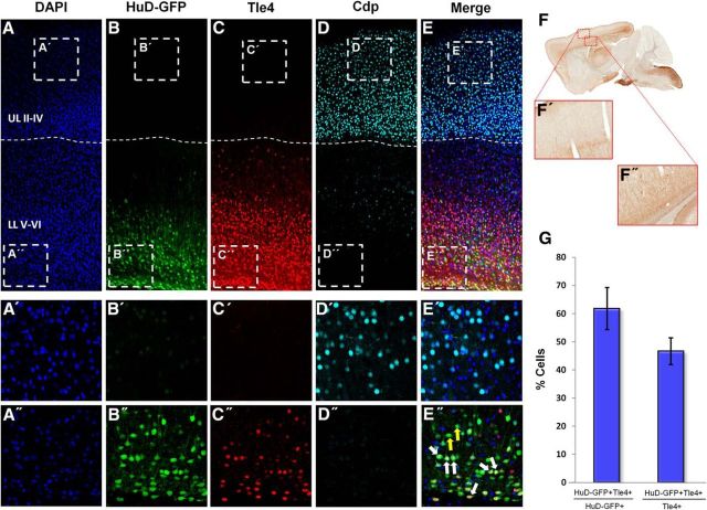
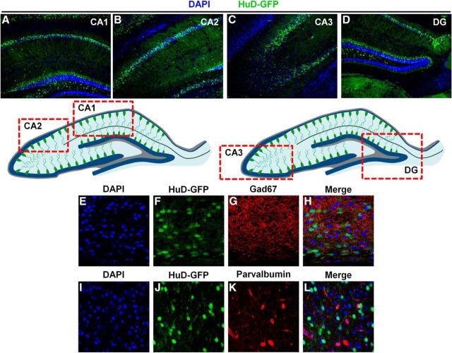
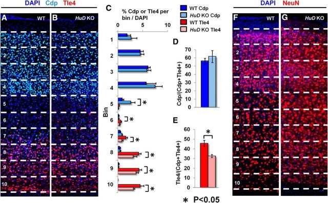
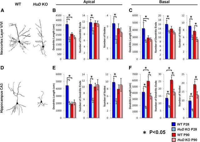
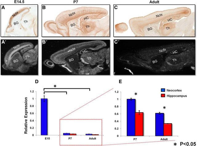
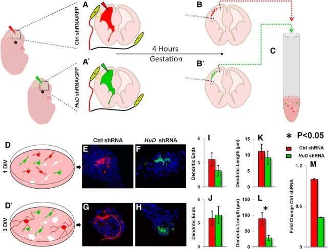
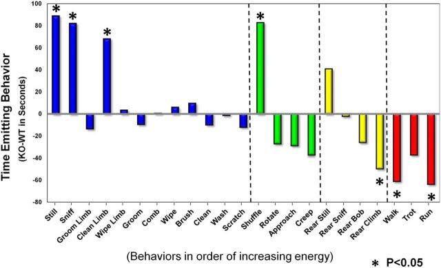
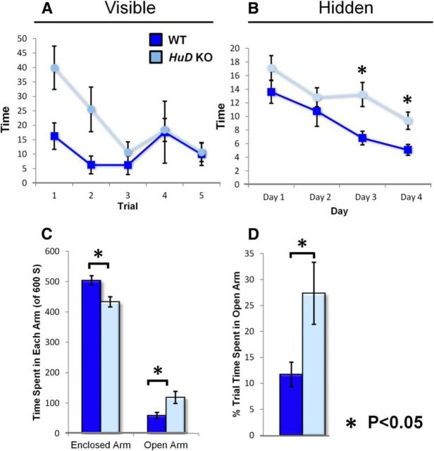
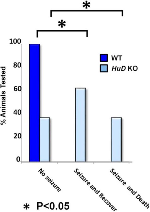
The proper functions of cortical circuits are dependent upon both appropriate neuronal subtype specification and their maturation to receive appropriate signaling. These events establish a balanced circuit that is important for learning, memory, emotion, and complex motor behaviors. Recent research points to mRNA metabolism as a key regulator of this development and maturation process. Hu antigen D (HuD), an RNA-binding protein, has been implicated in the establishment of neuronal identity and neurite outgrowth in vitro. Therefore, we investigated the role of HuD loss of function on neuron specification and dendritogenesis in vivo using a mouse model. We found that loss of HuD early in development results in a defective early dendritic overgrowth phase and pervasive deficits in neuron specification in the lower neocortical layers and defects in dendritogenesis in the CA3 region of the hippocampus. Subsequent behavioral analysis revealed a deficit in performance of a hippocampus-dependent task: the Morris water maze. Further, HuD knock-out (KO) mice exhibited lower levels of anxiety than their wild-type counterparts and were overall less active. Last, we found that HuD KO mice are more susceptible to auditory-induced seizures, often resulting in death. Our findings suggest that HuD is necessary for the establishment of neocortical and hippocampal circuitry and is critical for their function.


Figures

Figure 1.
Figure 1.
HuD-GFP is expressed in lower- but not upper-layer primary neurons of the mature neocortex. AE, Representative 10× confocal images of the neocortical wall for DAPI (blue), HuD-GFP (green), Tle4 (red), Cdp (light blue), and merged channels, respectively. Dashed line demarcates upper versus lower neocortical layers. UL, Upper layers; LL, lower layers. For A′–E′, insets are representative 60× confocal images of upper neocortical layers; for A′′–E′′, insets are representative 60× confocal images of lower neocortical layers. White arrows indicate HuD-GFP/Tle4+ neurons. Yellow arrows indicate HuD-GFP+/Tle4 neurons. F, F′, Representative light microscopy image of HuD-GFP mouse sagittal brain section using anti-GFP-DAB staining (Gong et al., 2003). G, Quantification of the proportion of HuD-GFP+ neurons colocalized with Tle4 (left) and Tle4+ neurons colocalized with HuD-GFP (right).
Figure 2.
Figure 2.
HuD is not expressed in Gad67 or parvalbumin+ interneurons and is expressed in the CA1–3 and dentate gyrus of the hippocampus. AD, Representative 10× confocal images of hippocampal subregions CA1, CA2, CA3, and dentate gyrus, respectively. Bottom, Schematic of HuD-GFP expression in the hippocampus. Red boxes denote regions where representative confocal images were captured. EH, Representative 60× confocal images of cortical of DAPI (blue), HuD-GFP (green), Gad67 (red), and merged channels, respectively. IL, Representative 60× confocal images of cortical of DAPI (blue), HuD-GFP (green), parvalbumin (red), and merged channels, respectively.
Figure 3.
Figure 3.
Early loss of HuD function disrupts the specification of lower layer neocortical primary neurons. A, B, Representative confocal images of the neocortical wall of adult WT and HuD KO at P90. Numbers and dashed lines denote 10 equal bins for analysis from layer II (bin 1) to the subplate (bin 10). DAPI is shown in dark blue, Tle4 in red, and Cdp in light blue. C, Quantification of the number of Cdp+ or Tle4+ cells in each bin/the number of DAPI+ cells in the column. Numbers are reported as the proportion of DAPI+ cells that are Cdp+ or Tle4+. Mean bin proportion compared between WT and KO for each bin. D, Quantification of the proportion of Cdp+ neurons from total labeled neurons (Cdp+Tle4). E, Quantification of the proportion of Tle4+ neurons from total labeled neurons. F, G, Representative confocal images of the cortical plate of adult WT and HuD KO as in A and B. DAPI is shown in dark blue and NeuN in red.
Figure 4.
Figure 4.
HuD loss of function disrupts dendritogenesis in deep neocortical layers and the CA3 region of the hippocampus. A, D, Representative tracings of lower layer neocortical primary neurons (A) and hippocampal CA3 pyramidal neurons (D). B, Quantification of apical dendrite length, dendritic ends, and nodes in lower layer neocortical neurons. C, Quantification of basal dendrite length, dendritic ends, and nodes in lower layer neocortical neurons. E, Quantification of apical dendrite length, dendritic ends, and nodes in CA3 pyramidal neurons. F, Quantification of basal dendrite length, dendritic ends, and nodes in CA3 pyramidal neurons.
Figure 5.
Figure 5.
HuD expression decreases from E14.5 until adult and becomes more regionally specific. AC, Representative images of HuD-GFP mouse sagittal brain sections stained with anti GFP DAB. E14.5, P7, and adult, respectively (Gong et al., 2003). A′–C′, Representative images of HuD ISH mouse sagittal brain sections at E14.5, P7, and adult, respectively (Magdaleno et al., 2006). D, E, qRT-PCR of developing neocortex at E15, P7, and adult. Hippocampus analysis at P7 and adult. Gapdh was used for normalization. Values were normalized to E15 neocortex in D. Values were normalized to P7 neocortex in E.
Figure 6.
Figure 6.
HuD controls the earliest stages of dendrite outgrowth. AB′, Schematic of In utero electroporation and dissociation of Ctrl shRNA/RFP (top) and HuD shRNA/GFP in E13.5 developing neocortex. Developing neocortices were electroporated at E13.5 with either Ctrl shRNA (RFP) or HuD shRNA (GFP). After 4 h, neocortices were dissociated and cultured. C, Schematic of dissociation of electroporated neocortices for primary cell culture. D, D′, Schematic of cell cultures taken at 1 and 3 DIV for analysis. E, F, Representative 60× confocal images of Ctrl and HuD shRNA transfected neurons at 1 DIV, respectively. G, H, Representative 60× confocal images of Ctrl and HuD shRNA transfected neurons at 3 DIV, respectively. IL, Quantification of neurite endings in 1 and 3 DIV cell cultures. M, qRT-PCR analysis of HuD shRNA efficiency in vitro. Gapdh was used as a normalization control.
Figure 7.
Figure 7.
Spectral analysis of HuD KO shows reduced overall activity and increased stereotypic behaviors. Spectral analysis of total time spent performing each behavior where HuD KO mean-WT mean for each. Analysis was subdivided into four main categories; stationary, orienting, rearing, and moving.
Figure 8.
Figure 8.
HuD KO mice perform poorly in Morris water maze and spend more time in the open arms of the elevated plus maze. A, Average latency to find a visible platform by genotype in five trials of the Morris water maze. B, Average latency to find a hidden platform by genotype in four consecutive days of testing, five trials per day. C, Average total time spent in the enclosed and open arms of the elevated plus maze by genotype. D, Proportion of total time spent in the open arms of the elevated plus maze by genotype.
Figure 9.
Figure 9.
HuD KO mice are more susceptible to auditory induced seizure than WT. Shown is a quantification of the proportion of mice that did not experience seizure, experienced a seizure and subsequently recovered, or experienced seizure and immediately died.

Similar articles

Cited by

References

    1. Abdelmohsen K, Hutchison ER, Lee EK, Kuwano Y, Kim MM, Masuda K, Srikantan S, Subaran SS, Marasa BS, Mattson MP, Gorospe M. miR-375 inhibits differentiation of neurites by lowering HuD levels. Mol Cell Biol. 2010;30:4197–4210. doi: 10.1128/MCB.00316-10. - DOI - PMC - PubMed
    1. Akamatsu W, Fujihara H, Mitsuhashi T, Yano M, Shibata S, Hayakawa Y, Okano HJ, Sakakibara S, Takano H, Takano T, Takahashi T, Noda T, Okano H. The RNA-binding protein HuD regulates neuronal cell identity and maturation. Proc Natl Acad Sci U S A. 2005;102:4625–4630. doi: 10.1073/pnas.0407523102. - DOI - PMC - PubMed
    1. Anderson GR, Galfin T, Xu W, Aoto J, Malenka RC, Südhof TC. Candidate autism gene screen identifies critical role for cell-adhesion molecule CASPR2 in dendritic arborization and spine development. Proc Natl Acad Sci U S A. 2012;109:18120–18125. doi: 10.1073/pnas.1216398109. - DOI - PMC - PubMed
    1. Anderson KD, Morin MA, Beckel-Mitchener A, Mobarak CD, Neve RL, Furneaux HM, Burry R, Perrone-Bizzozero NI. Overexpression of HuD, but not of its truncated form HuD I+II, promotes GAP-43 gene expression and neurite outgrowth in PC12 cells in the absence of nerve growth factor. J Neurochem. 2000;75:1103–1114. doi: 10.1046/j.1471-4159.2000.0751103.x. - DOI - PubMed
    1. Anderson KD, Sengupta J, Morin M, Neve RL, Valenzuela CF, Perrone-Bizzozero NI. Overexpression of HuD accelerates neurite outgrowth and increases GAP-43 mRNA expression in cortical neurons and retinoic acid-induced embryonic stem cells in vitro. Exp Neurol. 2001;168:250–258. doi: 10.1006/exnr.2000.7599. - DOI - PubMed

Publication types

MeSH terms