. 2014 May;165(1):52-61.
doi: 10.1104/pp.114.236539. Epub 2014 Mar 5.

Plants utilize a highly conserved system for repair of NADH and NADPH hydrates

Affiliations

Plants utilize a highly conserved system for repair of NADH and NADPH hydrates

Tom D Niehaus et al. Plant Physiol. 2014 May.

Abstract

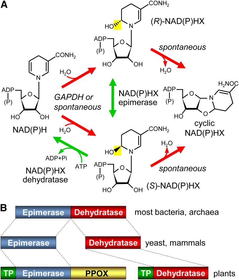
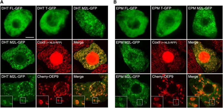
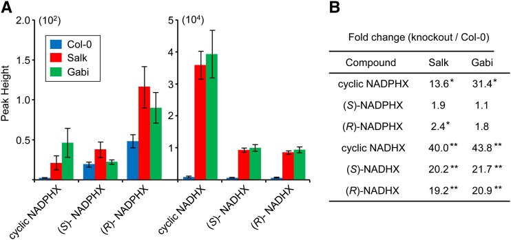
NADH and NADPH undergo spontaneous and enzymatic reactions that produce R and S forms of NAD(P)H hydrates [NAD(P)HX], which are not electron donors and inhibit various dehydrogenases. In bacteria, yeast (Saccharomyces cerevisiae), and mammals, these hydrates are repaired by the tandem action of an ADP- or ATP-dependent dehydratase that converts (S)-NAD(P)HX to NAD(P)H and an epimerase that facilitates interconversion of the R and S forms. Plants have homologs of both enzymes, the epimerase homolog being fused to the vitamin B6 salvage enzyme pyridoxine 5'-phosphate oxidase. Recombinant maize (Zea mays) and Arabidopsis (Arabidopsis thaliana) NAD(P)HX dehydratases (GRMZM5G840928, At5g19150) were able to reconvert (S)-NAD(P)HX to NAD(P)H in an ATP-dependent manner. Recombinant maize and Arabidopsis epimerases (GRMZM2G061988, At5g49970) rapidly interconverted (R)- and (S)-NAD(P)HX, as did a truncated form of the Arabidopsis epimerase lacking the pyridoxine 5'-phosphate oxidase domain. All plant NAD(P)HX dehydratase and epimerase sequences examined had predicted organellar targeting peptides with a potential second start codon whose use would eliminate the targeting peptide. In vitro transcription/translation assays confirmed that both start sites were used. Dual import assays with purified pea (Pisum sativum) chloroplasts and mitochondria, and subcellular localization of GFP fusion constructs in tobacco (Nicotiana tabacum) suspension cells, indicated mitochondrial, plastidial, and cytosolic localization of the Arabidopsis epimerase and dehydratase. Ablation of the Arabidopsis dehydratase gene raised seedling levels of all NADHX forms by 20- to 40-fold, and levels of one NADPHX form by 10- to 30-fold. We conclude that plants have a canonical two-enzyme NAD(P)HX repair system that is directed to three subcellular compartments via the use of alternative translation start sites.


Figures

Figure 1.
Figure 1.
NAD(P)H damage and repair reactions, and domain structures of the repair enzymes. A, The spontaneous or GAPDH-mediated hydration of NAD(P)H, and the enzymatic epimerization and dehydratase reactions that reconvert the resulting hydrates, (R)- and (S)-NAD(P)HX, to NAD(P)H. NAD(P)H hydrates can also spontaneously cyclize. The bacterial dehydratase reaction is ADP dependent rather than ATP dependent. B, Domain architectures of the NAD(P)HX epimerase and dehydratase enzymes of prokaryotes, yeast, and mammals, and of epimerase and dehydratase homologs in plants. TP, Predicted organellar targeting peptide.
Figure 2.
Figure 2.
In vitro activities of maize and Arabidopsis NAD(P)HX dehydratase and epimerase-PPOX proteins. A, Activities monitored spectrophotometrically at 22°C. Assays contained 25 mm Tris-HCl, pH 8.0, 5 mm KCl, 2 mm MgCl2, 0.1 mg/mL bovine serum albumin, 90 µm NADHX [55% (S)-NADHX, 45% (R)-NADHX], and 1 mm ATP (black circles) or ADP (white circles). Reactions were started by adding 5 µg of maize or Arabidopsis NAD(P)HX dehydratase and monitoring A340. At 5 min, 10 µg of the respective maize or Arabidopsis NAD(P)HX epimerase-PPOX (Epm) was added. Data are means ± se of three replicates; where no error bars appear, they are smaller than the symbols. B, Activities monitored by HPLC. Assays were as above but contained 1 mm NADHX, 5 mm ATP, and 20 µg of maize NAD(P)HX dehydratase (Dht) and epimerase-PPOX as indicated, and were incubated at 30°C for 30 min. A control without enzyme (no Enz) was included; the profile of this sample was the same as a sample given no incubation (not shown). Reactions were analyzed by HPLC, monitoring A279 (blue trace) and A340 nm (red trace). Peak fronts of (S)-NADHX, (R)-NADHX, and NADH are marked by dashed lines. Arabidopsis proteins gave similar results.
Figure 3.
Figure 3.
Epimerase activity of the epimerase domain, but not the PPOX domain, of the Arabidopsis NAD(P)HX epimerase-PPOX protein. Assays contained 25 mm Tris-HCl, pH 9.0, 1 mm KCl, NADHX preparations enriched in (S)-NADHX (1 mm; A) or (R)-NADHX (0.5 mm; B), and 10 µg of the separate epimerase (Epm) or PPOX domains. Incubation was at 30°C for 10 min. Controls without enzyme (no Enz) were included. Reactions were analyzed by HPLC, monitoring A279. Peak fronts of (S)- and (R)-NADHX are marked by dashed lines.
Figure 4.
Figure 4.
The in vitro translation products of Arabidopsis NAD(P)HX dehydratase and NAD(P)HX epimerase-PPOX, and their import into purified pea mitochondria and chloroplasts. A, Analysis of the products of in vitro transcription-translation. cDNAs that were full length (FL) or truncated to begin at the second predicted start Met (T), or with the second predicted start Met changed to Leu (M2L) were transcribed and translated in a wheat germ system containing [3H]Leu. The translation products were resolved by SDS-PAGE and visualized by fluorography. Positions of molecular mass markers (in kilodaltons) are indicated. The engineered (T and M2L) epimerase-PPOX constructs both give an additional minor translation product smaller than either of the others; this may reflect translation initiation at a third cryptic start site unmasked by the engineering. B, Dual import assays with pea chloroplasts and mitochondria. Dehydratase and epimerase-PPOX full-length sequences with the second Met changed to Leu (M2L), or truncated at the second Met (T), were translated in vitro as above. The translation products were incubated for 20 min in the light with mixed mitochondria (M) and chloroplasts (C), which were then reisolated using a Percoll gradient, without (−) or with (+) prior thermolysin treatment to remove adsorbed proteins. Proteins were separated by SDS-PAGE and visualized by fluorography. Samples were loaded next to an aliquot of the translation product (P).
Figure 5.
Figure 5.
Representative epifluorescence and confocal micrographs of tobacco BY-2 cells transiently expressing GFP fused to the C-terminus of either the native full-length sequence (FL), the truncated sequence beginning at the second predicted start site (T), or the full-length sequence with the second start site Met changed to Leu (M2L) of Arabidopsis NAD(P)HX dehydratase (DHT; A) or Arabidopsis NAD(P)HX epimerase/PPOX (EPM; B). The top rows are epifluorescence images that show, for both dehydratase and epimerase, the localization of the FL and T fusions in the cytosol and the localization of the M2L fusions predominantly to mitochondria. The middle rows are confocal images showing colocalization of the dehydratase and epimerase M2L fusions with endogenous mitochondrial cytochrome c oxidase subunit II. Note that these cells also exhibit red fluorescence in the nucleus because of the coexpressed NLS-RFP fusion protein, which was used as a convenient cell transformation marker. The bottom rows are confocal images showing the colocalization of the dehydratase and epimerase M2L fusions with the plastid (outer envelope) marker protein Cherry-OEP9 that was observed in approximately 5% to 10% of transformed cells. The boxes represent the portion of the cells shown at higher magnification in the insets. NLS-RFP, Nuclear localization signal fused to the red fluorescent protein. Bar = 10 μm in A.
Figure 6.
Figure 6.
Relative levels of pyridine nucleotide hydrates in Arabidopsis wild-type and NAD(P)HX dehydratase knockouts. Extracts of 14-d-old Arabidopsis wild-type (Col-0), Salk_203790 (Salk), or Gabi_173F11 (Gabi) seedlings (100 mg) were analyzed by LC-MS. A, Average peak areas of NADPHX forms (×102 scale) and NADHX forms (×104 scale) from six independent samples. B, The fold changes between knockouts and Col-0. *P < 0.05; **P < 0.01. Col-0, Ecotype Columbia 0 of Arabidopsis.

References

    1. Acheson SA, Kirkman HN, Wolfenden R. (1988) Equilibrium of 5,6-hydration of NADH and mechanism of ATP-dependent dehydration. Biochemistry 27: 7371–7375 - PubMed
    1. Bradford MM. (1976) A rapid and sensitive method for the quantitation of microgram quantities of protein utilizing the principle of protein-dye binding. Anal Biochem 72: 248–254 - PubMed
    1. Brandizzi F, Irons S, Kearns A, Hawes C. (2003) BY-2 cells: culture and transformation for live cell imaging. Curr Protoc Cell Biol Chapter 1: 1.7.1–1.7.16 - PubMed
    1. Breslow DK, Cameron DM, Collins SR, Schuldiner M, Stewart-Ornstein J, Newman HW, Braun S, Madhani HD, Krogan NJ, Weissman JS. (2008) A comprehensive strategy enabling high-resolution functional analysis of the yeast genome. Nat Methods 5: 711–718 - PMC - PubMed
    1. Chabregas SM, Luche DD, Van Sluys MA, Menck CF, Silva-Filho MC. (2003) Differential usage of two in-frame translational start codons regulates subcellular localization of Arabidopsis thaliana THI1. J Cell Sci 116: 285–291 - PubMed

Publication types

MeSH terms

LinkOut - more resources