. 2014 Apr 1;111(13):4970-5.
doi: 10.1073/pnas.1323279111. Epub 2014 Mar 5.

Rapid generation of a mouse model for Middle East respiratory syndrome

Affiliations

Rapid generation of a mouse model for Middle East respiratory syndrome

Jincun Zhao et al. Proc Natl Acad Sci U S A. .

Abstract

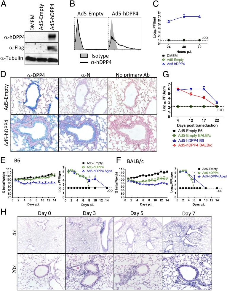
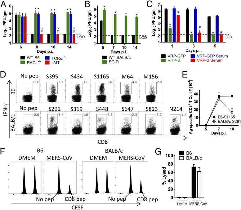
In this era of continued emergence of zoonotic virus infections, the rapid development of rodent models represents a critical barrier to public health preparedness, including the testing of antivirus therapy and vaccines. The Middle East respiratory syndrome coronavirus (MERS-CoV) was recently identified as the causative agent of a severe pneumonia. Given the ability of coronavirus to rapidly adapt to new hosts, a major public health concern is that MERS-CoV will further adapt to replication in humans, triggering a pandemic. No small-animal model for this infection is currently available, but studies suggest that virus entry factors can confer virus susceptibility. Here, we show that mice were sensitized to MERS-CoV infection by prior transduction with adenoviral vectors expressing the human host-cell receptor dipeptidyl peptidase 4. Mice developed a pneumonia characterized by extensive inflammatory-cell infiltration with virus clearance occurring 6-8 d after infection. Clinical disease and histopathological changes were more severe in the absence of type-I IFN signaling whereas the T-cell response was required for virus clearance. Using these mice, we demonstrated the efficacy of a therapeutic intervention (poly I:C) and a potential vaccine [Venezuelan equine encephalitis replicon particles expressing MERS-CoV spike protein]. We also found little protective cross-reactivity between MERS-CoV and the severe acute respiratory syndrome-CoV. Our results demonstrate that this system will be useful for MERS-CoV studies and for the rapid development of relevant animal models for emerging respiratory viral infections.

Keywords: SARS; emerging pathogen; interferon.


Conflict of interest statement

The authors declare no conflict of interest.

Figures

Fig. 1.
Fig. 1.
Development of mice susceptible to MERS-CoV infection. To assess hDPP4 expression (A) and surface localization (B), MLE15 cells were transduced with Ad5-hDPP4 or Ad5-Empty at an MOI of 20 at 37 °C for 4 h. hDDP4 expression was monitored by Western blot assay (A) or flow cytometry (B). (C) Ad5-hDPP4–transduced cells were infected with MERS-CoV at an MOI of 1 at 48 h posttransduction, and virus titers were determined by plaque assay. Five days after transduction with 2.5 × 10⁸ pfu of Ad5-hDPP4 or Ad5-Empty in 75 μL of DMEM intranasally, mice were intranasally infected with 1 × 10⁵ pfu of MERS-CoV in 50 μL of DMEM. (D) Lungs were harvested from BALB/c mice at day 3 after MERS-CoV infection, fixed in zinc formalin, and embedded in paraffin. Sections were stained with an anti-hDPP4 or with an anti–MERS-CoV nucleocapsid antibody (blue signal). (Original magnification, 20×.) (E and F) Weight changes in 6- to 12-wk-old (young) and 18- to 22-mo-old (aged) B6 (E) and BALB/c (F) mice were monitored daily. For B6 mice, n = 8 in Ad5-Empty group; 12 in Ad5-hDPP4 group; 8 in Ad5-hDPP4 aged group. For BALB/c mice, n = 8 in Ad5-Empty group; 12 in Ad5-hDPP4 group; 8 in Ad5-hDPP4 aged group. To obtain virus titers, lungs were homogenized at the indicated time points and titered on Vero 81 cells. Titers are expressed as pfu/g tissue (n = 4–8 mice per group per time point). Data are representative of two independent experiments. Δ, P < 0.05 when Ad5-hDPP4 aged were compared with Ad5-hDPP4 and Ad4-Empty. (G) To evaluate the length of time that Ad5-hDPP4–transduced mice could be infected with MERS-CoV, Ad5-hDPP4–transduced mice were infected with MERS-CoV at the indicated times. Lungs were harvested for titers at 2 d.p.i. (n = 4 mice per group per time point. (H) Lungs from B6 mice were removed at the indicated time points p.i., fixed in zinc formalin, and embedded in paraffin. Sections were stained with hematoxylin/eosin.
Fig. 2.
Fig. 2.
Requirements for type-I IFN induction and signaling in MERS-CoV clearance. (A) Five days after transduction with 2.5 × 10⁸ pfu of Ad5-hDPP4, mice were intranasally infected with 1 × 10⁵ pfu of MERS-CoV. Weight changes were monitored daily (n = 8 in B6 group; 14 in IFNAR−/− group; and 13 in MyD88−/− group; n= 9 in MAVS−/− group). (B) Virus titers in the lungs were measured at the indicated time points. Titers are expressed as pfu/g tissue (n = 3–4 mice per group per time point). Data are representative of two independent experiments. *, P < 0.05 compared with B6 group; Δ, P values of <0.05 compared with IFNAR−/− group; #, P values of <0.05 compared with MyD88−/− group. (C) Photographs of gross pathological lung specimens isolated from infected mice at day 7 p.i. (D) Sections of paraffin-embedded lungs from Ad5-hDPP4–transduced, infected IFNAR−/− mice were stained with hematoxylin/eosin. (E) Edema (asterisks) and infiltrating eosinophils (arrows) are indicated. (F) Ad5-hDPP4-–transduced mice were treated with 20 μg of poly I:C, 2,000 units of IFN-β, 200 ng of IFN-γ, or PBS in 50 μL of DMEM 6 h before intranasal infection with 1 × 10⁵ pfu of MERS-CoV. Viral titers in lungs were measured at the indicated time points (n = 4 mice per group per time point. Data are representative of three independent experiments. *, P < 0.05 compared with PBS group; Δ, P values of <0.05 compared with poly I:C group; #, P values of <0.05 compared with IFN-β group.
Fig. 3.
Fig. 3.
Requirements for CD8 T cells and antibodies for MERS-CoV clearance and protection from subsequent challenge. (A and B) Ad5-hDPP4–transduced mice were infected with 1 × 10⁵ pfu of MERS-CoV. Virus titers in the lungs were measured at the indicated time points. Titers are expressed as pfu/g tissue (n = 3–4 mice per group per time point. Data are representative of two independent experiments. *, P values of <0.05 compared with WT group; Δ, P values of <0.05 compared with RAG1−/− group; #, P values of <0.05 compared with TCRα−/− group. (C) BALB/c mice were immunized with 1 × 10⁵ infectious units (IU) of VRP-GFP or VRP-S in the footpad in 20 μL of PBS and boosted with the same dose 4 wk later. Mice were transduced and infected with 1 × 10⁵ pfu of MERS-CoV 2–4 wk after the booster. For adoptive transfer of serum, sera were obtained 2–4 wk after booster. Then, 300 μL of serum was transferred into transduced mice intraperitoneally 1 d before MERS-CoV infection. *, P values of <0.05 compared with VRP-GFP group; #, P values of <0.05 compared with VRP-GFP serum group. (D) To identify MERS-CoV–specific CD8 T-cell epitopes, single-cell suspensions were prepared from the lungs of transduced/infected mice and stimulated with 1 µM peptides for 5–6 h in the presence of brefeldin A. Frequencies of MERS-CoV–specific T cells (determined by IFN-γ intracellular staining) are shown. Kinetics of immune responses to dominant CD8 T-cell epitopes in Ad5-hDPP4–transduced B6 and BALB/c mice are summarized in E. Data are representative of five independent experiments. (F and G) In vivo cytotoxicity assays were performed on day 7 (BALB/c mice; peptides S395, S434, and S1165 combined) or day 8 (B6 mice; peptides S291, S823, and N214 combined) p.i. as described in Materials and Methods (F) and summarized (G) (n = 4 mice per group). Data are representative of two independent experiments.
Fig. 4.
Fig. 4.
Low level of cross-reactivity between MERS-CoV and SARS-CoV. (AC) Ad5-hDPP4–transduced BALB/c mice were inoculated with 1 × 10⁵ pfu of MERS-CoV or DMEM and rested for 5 wk before infection with 1 × 10⁴ pfu of SARS-CoV in 50 μL of DMEM. Control mice received 300 μL of immune serum from mice previously (5 wk) infected with a sublethal dose of SARS-CoV (500 pfu). Mortality (A) and weight (B) were monitored daily (n = 12 in all groups). (C) To obtain virus titers, lungs were homogenized at the indicated time points and titered on Vero E6 cells. Titers are expressed as pfu/g tissue (n = 4 mice per group per time point). Data are representative of two independent experiments. *, P values of <0.05 compared with DMEM group; Δ, P values of <0.05 compared with MERS-CoV–immunized group. (DF) BALB/c mice were infected with 500 pfu of SARS-CoV or DMEM and rested for 5 wk before Ad5-hDPP4 transduction and infection with 1 × 10⁵ pfu of MERS-CoV. Control mice received 300 μL of immune serum from mice previously (5 wk) infected with 1 × 10⁵ pfu of MERS-CoV. Mortality (D) and weight (E) were monitored daily (n = 12 in all groups). (F) Virus titers in the lungs were measured at the indicated time points (n = 4 mice per group per time point). Data are representative of two independent experiments. *, P values of <0.05 compared with DMEM group; Δ, P values of <0.05 compared with SARS-CoV–immunized group.

References

    1. Zaki AM, van Boheemen S, Bestebroer TM, Osterhaus AD, Fouchier RA. Isolation of a novel coronavirus from a man with pneumonia in Saudi Arabia. N Engl J Med. 2012;367(19):1814–1820. - PubMed
    1. Peiris JS, Guan Y, Yuen KY. Severe acute respiratory syndrome. Nat Med. 2004;10(Suppl 12):S88–S97. - PMC - PubMed
    1. Assiri A, et al. KSA MERS-CoV Investigation Team Hospital outbreak of Middle East respiratory syndrome coronavirus. N Engl J Med. 2013;369(5):407–416. - PMC - PubMed
    1. Assiri A, et al. Epidemiological, demographic, and clinical characteristics of 47 cases of Middle East respiratory syndrome coronavirus disease from Saudi Arabia: A descriptive study. Lancet Infect Dis. 2013;13(9):752–761. - PMC - PubMed
    1. Munster VJ, de Wit E, Feldmann H. Pneumonia from human coronavirus in a macaque model. N Engl J Med. 2013;368(16):1560–1562. - PMC - PubMed

Publication types

MeSH terms