. 2014 Apr 25;289(17):12016-12028.
doi: 10.1074/jbc.M113.530956. Epub 2014 Mar 5.

Endothelial Kruppel-like factor 4 regulates angiogenesis and the Notch signaling pathway

Affiliations

Endothelial Kruppel-like factor 4 regulates angiogenesis and the Notch signaling pathway

Andrew T Hale et al. J Biol Chem. .

Abstract

Regulation of endothelial cell biology by the Notch signaling pathway (Notch) is essential to vascular development, homeostasis, and sprouting angiogenesis. Although Notch determines cell fate and differentiation in a wide variety of cells, the molecular basis of upstream regulation of Notch remains poorly understood. Our group and others have implicated the Krüppel-like factor family of transcription factors as critical regulators of endothelial function. Here, we show that Krüppel-like factor 4 (KLF4) is a central regulator of sprouting angiogenesis via regulating Notch. Using a murine model in which KLF4 is overexpressed exclusively in the endothelium, we found that sustained expression of KLF4 promotes ineffective angiogenesis leading to diminished tumor growth independent of endothelial cell proliferation or cell cycling effects. These tumors feature increased vessel density yet are hypoperfused, leading to tumor hypoxia. Mechanistically, we show that KLF4 differentially regulates expression of Notch receptors, ligands, and target genes. We also demonstrate that KLF4 limits cleavage-mediated activation of Notch1. Finally, we rescue Notch target gene expression and the KLF4 sprouting angiogenesis phenotype by supplementation of DLL4 recombinant protein. Identification of this hitherto undiscovered role of KLF4 implicates this transcription factor as a critical regulator of Notch, tumor angiogenesis, and sprouting angiogenesis.

Keywords: ATPases; Actin; Arabidopsis; Kinetics; Molecular Motors; Myosin.


Figures

FIGURE 1.
FIGURE 1.
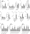
KLF4 has angiogenic effects. We designed human- and mouse-specific KLF4 primers to compare levels of overexpressed mKLF4 to endogenous human (hKLF4) as well as hKLF4 levels after siRNA-mediated knockdown. Total KLF4 (mKLF4 plus hKLF4) is expressed as fold-change from endogenous (hKLF4) alone (A) or by copy number (B). Data shown represents data pooled from HUVEC derived from 4 umbilical cords, normalized to succinate dehydrogenase (SDHA), and presented as mean ± S.E. C, KLF4 alters EC expression of several genes involved in angiogenesis. HUVEC were transduced with a Lenti-K4 or EV control and assessed by qPCR (C, left panel). In separate experiments, HUVEC were transfected with K4−/− siRNA or NS control (C, right panel). D, left panel, overexpression of KLF4 up-regulates VEGFA expression independent of hypoxia (norm, normoxia; hyp, hypoxia). D, right panel, knockdown of KLF4 results in decreased VEGFA expression independent of hypoxia. Gene expression is normalized to β-actin. E, commercially obtained EC were plated and exposed to hypoxia (3% O2) for the indicated number of hours, then harvested and assessed for hKLF4 expression by qPCR. F, ChIP experiments using an anti-KLF4 antibody versus IgG control showed enrichment of KLF4 at specific CACCC sites in the VEGFA, VEGFR1 and VEGFR2 promoters. Data shown represents data pooled from HUVEC derived from 3 cords (n = 3). 18 S is the non-template control. *, indicates p < 0.05 by Student's t test, and data are represented as mean ± S.E.
FIGURE 2.
FIGURE 2.
Overexpression of KLF4 regulates sprouting angiogenesis in vitro. A, KLF4 promotes EC contribution to sprout formation (cellular area) in a Matrigel model of sprouting angiogenesis (norm, normoxia; hyp, hypoxia). B, KLF4 overexpression causes an increase in total number of sprouts and number of sprout nodes, but does not alter individual sprout length or sprout:node ratio compared with EV HUVEC. C, KLF4 promotes EC contribution to sprout formation (cellular area) in GFR Matrigel. D, KLF4 overexpression causes an increase in the total number of sprouts and number of sprout nodes, but does not alter individual sprout length or sprout:node ratio in GFR Matrigel. E, representative images (×40) of HUVEC seeded and grown in Matrigel for ∼6–8 h. Data are representative of 9 wells per condition repeated three times (n = 3). *, p < 0.05 by ANOVA; **, p < 0.05 by Student's t test for paired normoxia-hypoxia samples, i.e. EV normoxia versus EV hypoxia). Data are presented as mean ± S.E. and represents HUVEC derived from 4–6 cords (n = 4–6). Images were quantified using ImageJ.
FIGURE 3.
FIGURE 3.
Knockdown of KLF4 regulates sprouting angiogenesis in vitro. A, decreased expression of KLF4 inhibits EC contribution to sprout formation. B, knockdown of KLF4 causes a decreased number of sprouts and nodes, but length/sprout and sprout:node ratio remains unchanged compared with NS HUVEC. C, KLF4 deficiency inhibits EC contribution to sprout formation in GFR Matrigel. D, knockdown of KLF4 decreased the number of sprouts per field, length per sprout, number of nodes, and sprout:node ratio in GFR Matrigel. E, representative images (×40) of HUVEC seeded and grown in Matrigel for ∼6–8 h. Data are representative of 9 wells per condition repeated three times (n = 3). *, p < 0.05 by ANOVA; **, p < 0.05 by Student's t test for paired normoxia-hypoxia samples, i.e. NS normoxia versus NS hypoxia). Data are presented as mean ± S.E. and represents HUVEC derived from 4–6 cords (n = 4–6). Images were quantified using ImageJ.
FIGURE 4.
FIGURE 4.
Sustained KLF4 overexpression causes ineffective sprouting angiogenesis. A, EC-K4 Tg tumors are significantly smaller than WT (1 × 10⁶ cells, left panel; 2.5 × 10⁶ cells, right panel). B, B16-F10 tumors were harvested 14 days after subcutaneous flank inoculation with 1 × 10⁶ melanoma cells. Tumors from 1 of 5 experiments (n = 4–12 animals per group; 1 × 10⁶ cells) are shown. C, IHC analysis demonstrates enhanced EC KLF4 signal (red, KLF4; green, PECAM; ×400). The vascular tumor rim was partitioned from harvested tumors and subjected to gene expression analysis. Sustained overexpression of human KLF4 was expressed as total KLF4 (mKLF4 plus hKLF4) and are represented as fold-change compared with WT. D, PECAM staining demonstrates significantly increased numbers of vessels in EC-K4 Tg tumors (×100, left; ×200, right). Quantification was performed on representative sections from 3 to 4 tumors per group (20–32 fields/tumor) and normalized to WT vessel density. E, EC-K4 Tg tumor rim homogenate has increased expression of angiogenesis-associated gene targets. WT and EC-K4 Tg tumor rim was harvested and gene expression was assessed and normalized to 36B4. F, vessels in EC-K4 Tg tumors are hypoperfused compared with WT tumor vessels. All vessels are stained red (PECAM), and only perfused vessels are labeled with FITC-lectin (green). Complete sections (7–8 fields) of each tumor with 4 tumors (850–900 vessels) per group were analyzed p < 0.025, ×200). G, intraperitoneal injection of Hypoxyprobe 1 h prior to tumor harvest results in labeling of proteins modified by exposure to a hypoxic environment. Distance from vascular structures of the tumor rim (PECAM, green) to hypoxic regions (red, outlined in white) is decreased in EC-K4 Tg tumors (n = 8 sections per group; AU, arbitrary units). H, we observed no gross difference of melanoma proliferation between WT and EC-K4 Tg tumors. Proliferation was assessed using IHC and staining for proliferating cell nuclear antigen (green) and DAPI (blue) (×100). Quantification of PCNA staining is shown by the ratio of PCNA-positive cells to total number of cells (DAPI+) per field (p = NS; 5–10 sections per tumor and 4 tumors were analyzed). *, indicates location of the outer tumor surface. I, the vascular tumor rim was partitioned and subjected to gene expression analysis. Data are normalized to 36b4 and represents 4 tumors per group (n = 4) presented as mean ± S.E. J, no gross difference in pericyte coverage between WT and EC KLF4 Tg tumor vessels was observed. Assessment of pericyte coverage was performed by IHC using PECAM (green) and NG-2 or laminin (red) staining. Representative photographs with PECAM and laminin co-staining are shown (]times]200; p = NS, 6–7 sections per tumor and 3–4 tumors per group were analyzed). Data are presented as mean ± S.E.
FIGURE 5.
FIGURE 5.
The role of KLF4 in EC proliferation, apoptosis, migration, and metabolism. A, lentiviral overexpression of KLF4 does not affect EC proliferation as assessed by cell count quantification over a 3-day incubation period (n = 4 independent experiments, 12 total wells per group). B, sustained overexpression of KLF4 does not alter Caspase 3/7-activity under either basal conditions or with hypoxia stimulation (n = 5 independent preps per group). C, overexpression of KLF4 does not alter EC migration after “wound induction” (scratch assay, n = 3 independent experiments, nine total wells per group). D, metabolism (alamarBlue assay, n = 3–4 independent experiments, 18–24 total wells per group) is unchanged with overexpression of KLF4. alamarBlue assay measures redox changes of the growth medium, indicative of metabolic activity. Data are presented as mean ± S.E. E, knockdown of KLF4 inhibits EC proliferation. *, p < 0.05 by Student's t test for paired samples, i.e. day 1 NS versus day 1 K4−/−. F, knockdown of KLF4 increases Caspase 3/7 activity. *, p < 0.05 by ANOVA; **, p < 0.05 by Student's t test for paired normoxia-hypoxia samples, i.e. K4−/− normoxia versus K4−/− hypoxia. G, cell migration is inhibited with knockdown of KLF4. *, p < 0.05 by ANOVA. H, metabolism is decreased with knockdown of KLF4. *, p < 0.05 by Student's t test. Data are presented as mean ± S.E. and represents HUVEC derived from 4–6 cords (n = 4–6).
FIGURE 6.
FIGURE 6.
KLF4 overexpression does not effect EC cell cycling. A, overexpression of KLF4 does not affect EC cell cycle regulation as shown by flow cytometric analysis using BrdU and 7-amino-actinomycin D (7-ADD). One representative dot plot per group is shown. B, quantification for R2 (G0/1 transition), R3 (S phase), R4 (G2/M transition), and R5 (apoptosis) are shown as pooled data (n = 2 independent experiments, 3 EC preps per experiment). C, overexpression of KLF4 in HUVEC does not alter expression of 18 cell cycle-associated genes. Data are presented as mean ± S.E. from 3 independent experiments, normalized to expression in control (EV) samples. *, indicates p < 0.05.
FIGURE 7.
FIGURE 7.
KLF4 regulates Notch expression. A, KLF4 differentially regulates Notch family member expression in EC-K4 Tg tumors. Tumor rim homogenate was harvested and assessed for candidate gene expression. Data are normalized to PECAM, presented as mean ± S.E. and is pooled from 3–5 tumors per group (n = 3–5). KLF4 regulates Notch receptor (B, left panel), ligand (C, left panel), and target gene (D, left panel) mRNA expression levels in HUVEC. Control samples are normalized to 1 and fold-change values are expressed relative to the respective control. Data are presented as mean ± S.E., and represents HUVEC derived from 4–6 cords (n = 4–6). B–D, right panels, KLF4 differentially regulates Notch family member mRNA expression after Notch activation by NICD. Endogenous gene expression was assessed after infection with KLF4- or NICD-expressing virus and respective control virus. (For E–G, commercially available HUVEC were used, n = 3–4 independent experiments, *, p < 0.05.) E, KLF4 inhibits CSL-mediated Notch activation. A CBX4 reporter plasmid (concatemer with four CSL binding sites), KLF4 expression plasmid, and NICD-expressing virus were transfected into HUVEC and CBX4 activation was expressed as relative luciferase units (RLU). F, KLF4 inhibits Notch-mediated transcription of target gene HES1. HUVEC were transfected with a Hes1 promoter-reporter construct and KLF4 expression plasmid, NICD or both. G, Notch activation via NICD decreases KLF4 promoter activity (RLU). HUVEC were transfected with a KLF4 promoter-reporter construct and NICD expression plasmids. H, KLF4 is enriched on the CACCC sites on the promoters of NOTCH1, DLL4, and HES1 as assessed by ChIP in HUVEC. Data are derived from 3 separate cords and are normalized to input DNA. 18 S is the non-targeting DNA control. *, p < 0.05 by Student's t test.
FIGURE 8.
FIGURE 8.
KLF4 negatively regulates Notch activation through NOTCH1 and DLL4. A, overexpression of KLF4 decreases production of NICD, the cleaved intracellular fragment of Notch1, in HUVEC. NICD signal is normalized to β-actin. γ-Secretase inhibitor and VEGFA treatment were used as negative and positive controls, respectively. One representative blot is shown. Densitometry analysis represents data pooled from four independent experiments (n = 4). B, inhibition of Notch target genes HES1, HEY1, and HEY2 by sustained KLF4 overexpression is reversed by supplementation of cultures with DLL4 recombinant protein. Data are presented as mean ± S.E. and represents HUVEC derived from 4 cords (n = 4). C, enhanced sprout formation in primary EC-K4 Tg spheroids is reversed by addition of DLL4 recombinant protein (EC were derived from 5 mice and pooled (n = 5), sprouts from 50 spheroids were counted per condition, *, p < 0.05 by ANOVA). Representative images (×4) of primary cardiac EC after growing 3–4 days in diluted Matrigel are shown in D.

References

    1. Iso T., Hamamori Y., Kedes L. (2003) Notch signaling in vascular development. Arterioscler. Thromb. Vasc. Biol. 23, 543–553 - PubMed
    1. Krebs L. T., Xue Y., Norton C. R., Shutter J. R., Maguire M., Sundberg J. P., Gallahan D., Closson V., Kitajewski J., Callahan R., Smith G. H., Stark K. L., Gridley T. (2000) Notch signaling is essential for vascular morphogenesis in mice. Genes Dev. 14, 1343–1352 - PMC - PubMed
    1. Hrabĕ de Angelis M., McIntyre J., 2nd, Gossler A. (1997) Maintenance of somite borders in mice requires the Delta homologue DLL1. Nature 386, 717–721 - PubMed
    1. Roca C., Adams R. H. (2007) Regulation of vascular morphogenesis by Notch signaling. Genes Dev. 21, 2511–2524 - PubMed
    1. Xue Y., Gao X., Lindsell C. E., Norton C. R., Chang B., Hicks C., Gendron-Maguire M., Rand E. B., Weinmaster G., Gridley T. (1999) Embryonic lethality and vascular defects in mice lacking the Notch ligand Jagged1. Hum. Mol. Genet. 8, 723–730 - PubMed

Publication types

LinkOut - more resources