. 2014 Apr 18;289(16):11083-11094.
doi: 10.1074/jbc.M114.557306. Epub 2014 Mar 5.

Structure-function analysis of Staphylococcus aureus amidase reveals the determinants of peptidoglycan recognition and cleavage

Affiliations

Structure-function analysis of Staphylococcus aureus amidase reveals the determinants of peptidoglycan recognition and cleavage

Felix Michael Büttner et al. J Biol Chem. .

Abstract

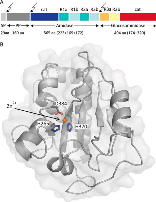
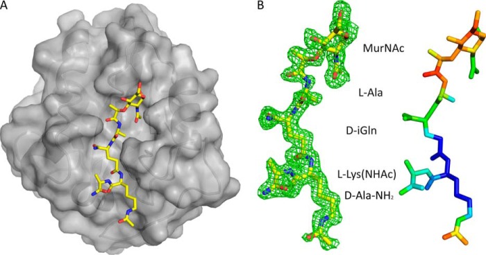
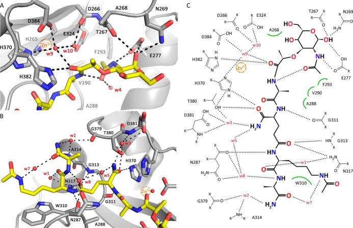
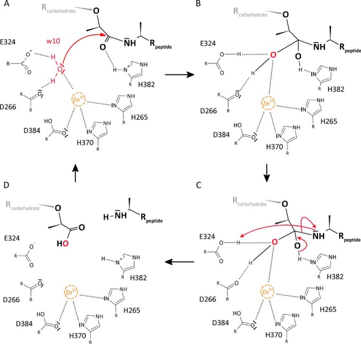
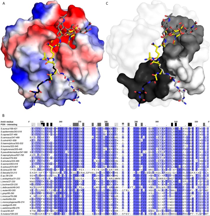
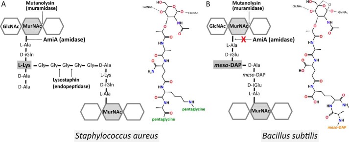
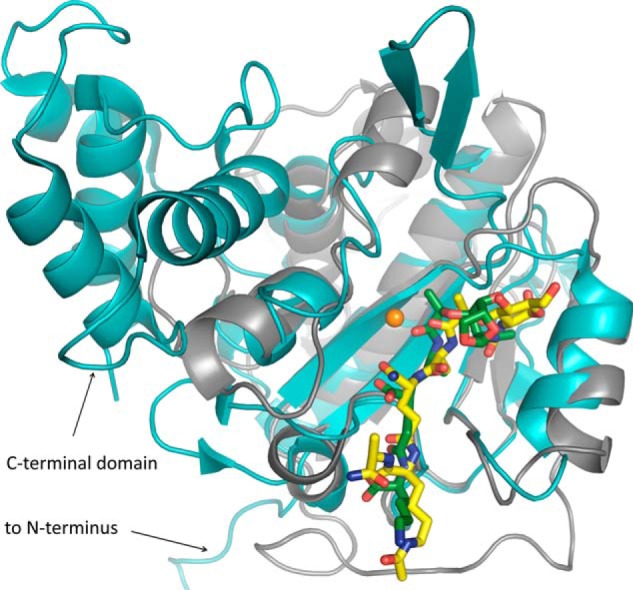
The bifunctional major autolysin AtlA of Staphylococcus aureus cleaves the bacterium's peptidoglycan network (PGN) at two distinct sites during cell division. Deletion of the enzyme results in large cell clusters with disordered division patterns, indicating that AtlA could be a promising target for the development of new antibiotics. One of the two functions of AtlA is performed by the N-acetylmuramyl-l-alanine amidase AmiA, which cleaves the bond between the carbohydrate and the peptide moieties of PGN. To establish the structural requirements of PGN recognition and the enzymatic mechanism of cleavage, we solved the crystal structure of the catalytic domain of AmiA (AmiA-cat) in complex with a peptidoglycan-derived ligand at 1.55 Å resolution. The peptide stem is clearly visible in the structure, forming extensive contacts with protein residues by docking into an elongated groove. Less well defined electron density and the analysis of surface features indicate likely positions of the carbohydrate backbone and the pentaglycine bridge. Substrate specificity analysis supports the importance of the pentaglycine bridge for fitting into the binding cleft of AmiA-cat. PGN of S. aureus with l-lysine tethered with d-alanine via a pentaglycine bridge is completely hydrolyzed, whereas PGN of Bacillus subtilis with meso-diaminopimelic acid directly tethered with d-alanine is not hydrolyzed. An active site mutant, H370A, of AmiA-cat was completely inactive, providing further support for the proposed catalytic mechanism of AmiA. The structure reported here is not only the first of any bacterial amidase in which both the PGN component and the water molecule that carries out the nucleophilic attack on the carbonyl carbon of the scissile bond are present; it is also the first peptidoglycan amidase complex structure of an important human pathogen.

Keywords: Amidase; Autolysin; Complex Structure; Drug Design; Enzyme Mechanisms; MRSA; Peptidoglycan; Staphylococcus aureus; X-ray Crystallography; Zinc.


Figures

FIGURE 1.
FIGURE 1.
Prepro-AtlA holoenzyme and structure of AmiA-cat. A, domain arrangement of AtlA with sites of post-translational cleavage indicated by arrows. SP, signal peptide; PP, propeptide; cat, catalytic domain; R, repeat domain. This figure was adapted and modified from Ref. . B, mixed α/β fold of unliganded AmiA-cat in a schematic representation with a transparent surface. The zinc ion (orange) and its coordinating residues (dark gray) are highlighted.
FIGURE 2.
FIGURE 2.
AmiA-cat in complex with MtetP. A, AmiA-cat (semitransparent gray surface) bound MtetP (yellow sticks) in the active site. B, the close-up on MtetP surrounded by omit density illustrates that the ligand is well defined. Still, the B-factor distribution of MtetP displays the elevated flexibility of its MurNAc moiety. The difference omit map is shown at a σ level of 2.0, and the color scale for B-factors ranges from blue (20 Å2) to red (50 Å2).
FIGURE 3.
FIGURE 3.
Interactions between AmiA-cat and MtetP. A, interactions of AmiA-cat with the MurNAc moiety and l-Ala of MtetP at the active site. The zinc ion from the native structure (orange sphere) is superimposed on the AmiA-cat complex (gray) with MtetP (yellow). B, close-up of the interactions of the peptide moiety of MtetP with AmiA. C, ChemSketch (57) plot of interactions between MtetP and AmiA-cat. Van-der-Waals interactions are depicted as green arcs, and hydrogen bonds are shown as black dashed lines. Coordination of Wat-9 involves His-370, Asp-384, and Wat-10 and a hydrogen bond with the carbonyl oxygen of the scissile amide bond in the ligand, which itself is positioned by interaction with His-382. Wat-10 lies next to Asp-266, Glu-324, Wat-9, and the carbonyl carbon of the scissile bond. MurNAc forms an intramolecular and four further hydrogen bonds with Glu-277, Thr-267, and Asn-269. Hydrophobic interactions of the methyl groups involve Ala-268 and Phe-293, respectively. l-Ala inserts into a small hydrophobic pocket formed by residues Ala-288 and Val-290. The amide side chain of d-iGln forms direct hydrogen bonds with Thr-380 and His-370 as well as two water-mediated hydrogen bonds to Asp-381. The carbonyl oxygen bonds with Gly-313 and Asn-317. l-Lys is stabilized by interactions with Asn-287 as well as Asn-317, whereas the acetylated side chain engages in hydrophobic interactions with Trp-310. A water (Wat-7 (w7)) bridges Nϵ of l-Lys-NHAc and the carbonyl oxygen of d-Ala, which engages additional water-mediated interactions with AmiA-cat residues Asn-287, Ala-314, and Gly-379.
FIGURE 4.
FIGURE 4.
Proposed reaction mechanism of AmiA. A, Wat-10 (w10) is hydrogen-bonded to Asp-266 and Glu-324, and its free electron pairs face toward the scissile bond. Zn2+ is complexed by His-265, His-370, and Asp-384 and probably renders Wat-10 more reactive, enabling a nucleophilic attack. B, tetrahedral intermediate is stabilized by hydrogen bonds of the resulting hydroxyl groups with Nδ of His-382 as well as Asp-266, Glu-324, and zinc, respectively. C, reformation of a carbonyl group with the peptide moiety as leaving group. His-382 can accept a hydrogen atom from the tetrahedral intermediate, whereas the peptide is poised to accept a hydrogen from Glu-324. D, product release.
FIGURE 5.
FIGURE 5.
AmiA-cat binding to PGN. A, electrostatic surface of AmiA-cat with MtetP (yellow), zinc (orange), and modeled PGN components (dark gray, black, and light gray). Uncharged (white surface area), positively charged (blue areas), and negatively charged residues (red surface) are shown. The spacious hydrophilic pocket harboring the zinc ion and active site also accommodates MurNAc. Adjacent GlcNAc rings (dark gray sticks) shift MurNAc slightly when modeled as a polymer. The lower peptide moiety of MtetP binds in the mostly uncharged region of the binding cleft. Two conformations for the pentaglycine bridge (black and light gray sticks, respectively) linked to l-Lys of MtetP were modeled according to uncharged surface area and possible hydrogen bonds. B, a multisequence alignment of bacterial amidases with identical residues colored by conservation from light to dark blue. Residues forming the carbohydrate binding pocket, marked by dark gray boxes, are highly conserved among all compared amidases. Surface-exposed amino acids near the two pentaglycine bridges are marked in black and light gray for the respective conformation models. Conservation, especially among staphylococci is high for both. Alignment was calculated using Clustal Omega (58), and the output was created using Jalview (59). C, conserved residues mapped on the AmiA-cat surface according to the color scheme used in A and B.
FIGURE 6.
FIGURE 6.
RP-HPLC profile of mutanolysin-digested PGN fragments after incubation with enzyme. A, S. aureus PGN fragments incubated with elution buffer of AmiA-cat as control. Three peaks around 28 min contain monomer species of one peptide stem with carbohydrates, peaks at 58 min comprise species with two peptide stems, peaks at 73 min cover fragments with three peptide stems, and continuing accordingly. B, S. aureus PGN fragments incubated with AmiA-cat result in completely digested polymers with GlcNAc-MurNAc fragments remaining. C, S. aureus PGN fragments incubated with active site mutant AmiA-H370A show no catalytic activity. D, B. subtilis PGN fragments incubated with elution buffer of AmiA-cat result in a pattern similar to A but with specific retention times for B. subtilis. E, B. subtilis PGN fragments incubated with AmiA-cat exhibit no activity of the staphylococcal amidase for B. subtilis PGN. F, mass analysis of the major product shown in B, which corresponds to the disaccharide GlcNAc-MurNAc (m/z 496).
FIGURE 7.
FIGURE 7.
Schematic and molecular illustration of the PGN structures. A, in S. aureus, the PGN subunit is composed of d-isoglutamine and l-lysine and is cross-linked via a pentaglycine bridge (43). It comprises C-terminally either d-Ala-d-Ala or d-Ala-pentaglycine. B, B. subtilis PGN from vegetative cells (53, 55), however, contains d-isoglutamic acid and amidated meso-DAP and lacks a d-alanine at the meso-DAP α-carboxyl group, and it is directly cross-linked with d-alanine of the next subunit. In addition, B. subtilis PGN has 1,6-anhydro-MurNAc, shown in gray, at its terminus instead of a reducing MurNAc.
FIGURE 8.
FIGURE 8.
Comparison of the AmiA-cat complex (gray schematic) with AmiD from E. coli (cyan schematic; Protein Data Bank code 3D2Y). Superimposition of the two bacterial amidases reveals a similar fold solely around the binding and active site. AmiD deviates by a 2.0-Å root mean square deviation (DaliLite pairwise) from the AmiA-cat main chain, amino acids in the active and binding sites differ, and AmiD contains additional motifs at its N and C termini. Ligand positioning of anhydro-MTP (green sticks) to AmiD is comparable with the AmiA-cat complex with MtetP (yellow sticks). However, anhydro-MTP has an overall shift in relation to MtetP, and interactions of enzyme with ligand are unalike. Additionally, the MurNAc moiety, including the scissile bond, lies in the direct vicinity of the zinc binding residues and is in the anhydro form, which does not occur in staphylococci. Zn2+ from the unliganded AmiA-cat structure in orange was superimposed.

References

    1. Baron S. (1996) in Medical Microbiology (Baron S., ed) 4th Ed., Chapter 12, University of Texas, Galveston, TX - PubMed
    1. O'Gara J. P., Humphreys H. (2001) Staphylococcus epidermidis biofilms: importance and implications. J. Med. Microbiol. 50, 582–587 - PubMed
    1. Götz F. (2002) Staphylococcus and biofilms. Mol. Microbiol. 43, 1367–1378 - PubMed
    1. Heilmann C., Schweitzer O., Gerke C., Vanittanakom N., Mack D., Götz F. (1996) Molecular basis of intercellular adhesion in the biofilm-forming Staphylococcus epidermidis. Mol. Microbiol. 20, 1083–1091 - PubMed
    1. Götz F. G. P. (2000) in Infections Associated with Indwelling Medical Devices (Waldvogel F. A., Bisno A. L., eds) pp. 55–88, American Society for Microbiology Press, Washington, D. C

Publication types

MeSH terms

LinkOut - more resources