. 2014 Jan 23:2014:592450.
doi: 10.1155/2014/592450. eCollection 2014.

p104 binds to Rac1 and reduces its activity during myotube differentiation of C2C12 cell

Affiliations

p104 binds to Rac1 and reduces its activity during myotube differentiation of C2C12 cell

Ki Young Choi et al. ScientificWorldJournal. .

Abstract

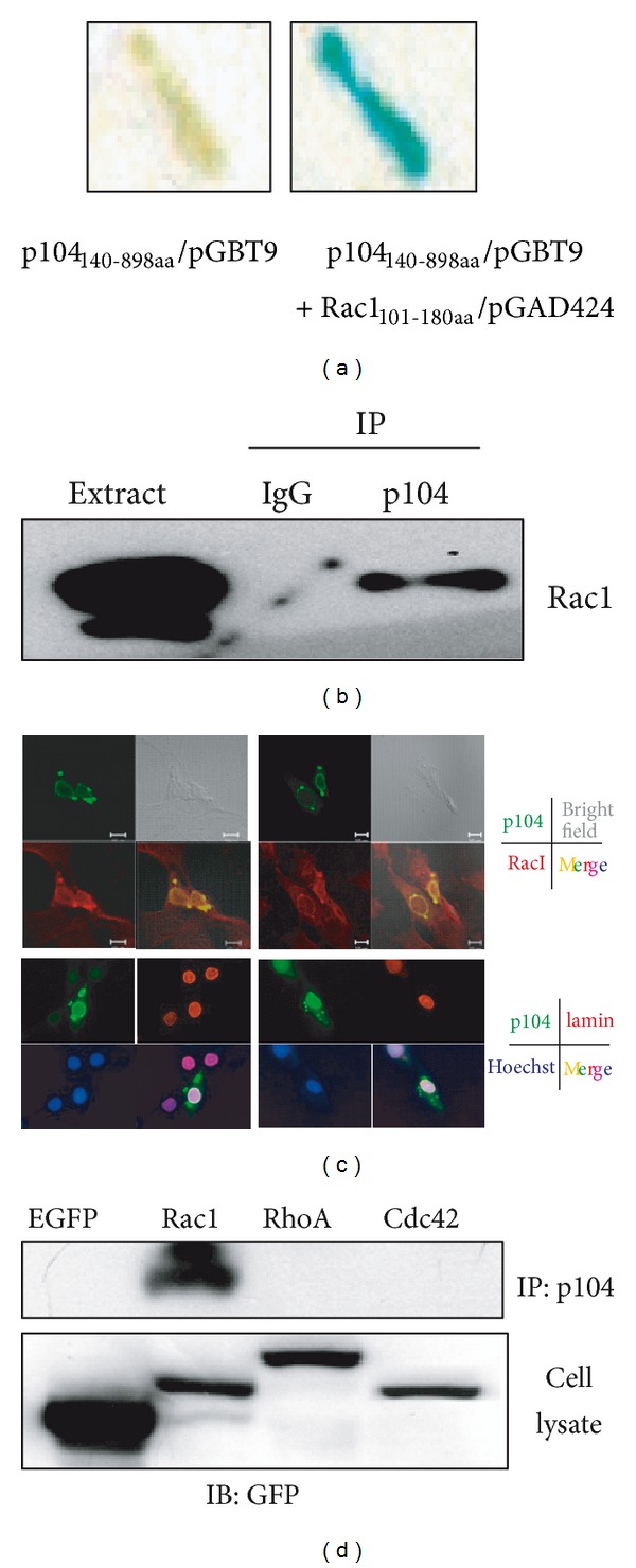
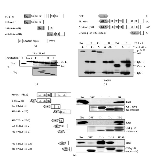
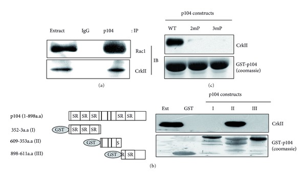
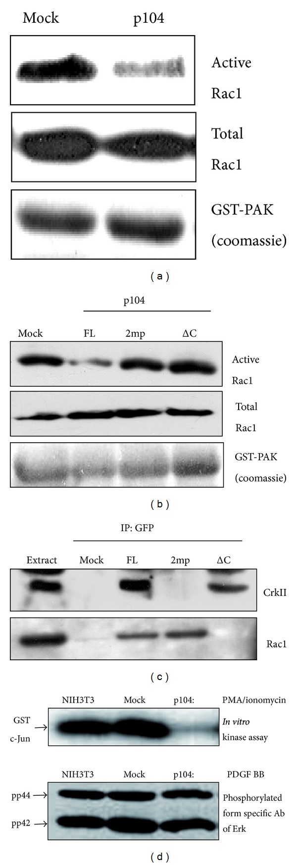
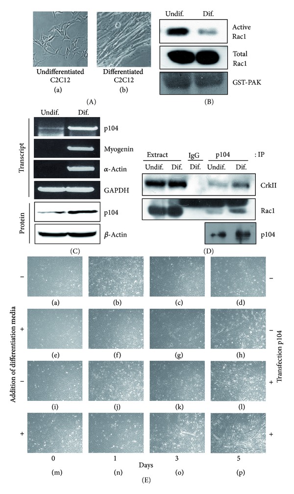
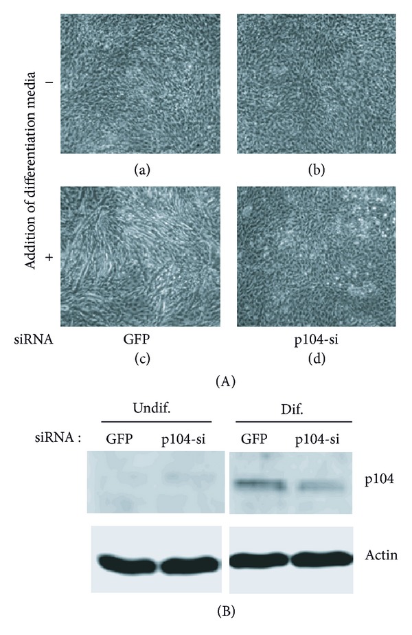
The p104 protein inhibits cellular proliferation when overexpressed in NIH3T3 cells and has been shown to associate with p85α, Grb2, and PLCγ1. In order to isolate other proteins that interact with p104, yeast two-hybrid screening was performed. Rac1 was identified as a binding partner of p104 and the interaction between p104 and Rac1 was confirmed by immunoprecipitation. Using a glutathione S-transferase (GST) pull-down assay with various p104 fragments, the 814-848 amino acid residue at the carboxyl-terminal region of p104 was identified as the key component to interact with Rac1. The CrkII which is involved in the Rac1-mediated cellular response was also found to interact with p104 protein. NIH3T3 cells which overexpressed p104 showed a decrease of Rac1 activity. However, neither the proline-rich domain mutant, which is unable to interact with CrkII, nor the carboxy-terminal deletion mutant could attenuate Rac1 activity. During the differentiation of myoblasts, the amount of p104 protein as well as transcript level was increased. The overexpression of p104 enhanced myotube differentiation, whereas siRNA of p104 reversed this process. In this process, more Rac1 and CrkII were bound to increased p104. Based on these results, we conclude that p104 is involved in muscle cell differentiation by modulating the Rac1 activity.


Figures

Figure 1
Figure 1
Rac1 was isolated as a binding protein of p104. (a) The DNA-encoding p104 (amino acids residue 140–898) was inserted into the EcoRI/Pst Isite of a GAL4 DNA-binding domain vector pGBT9. After colony selection on plates lacking Trp, His, and Leu, β-galactosidase assay was performed on the filter. Rac1 clones were isolated as a binding partner of p104. (b) Using anti-p104 serum, immunoprecipitation was performed with mouse brain extract and analyzed by Western blotting with an anti-Rac1 antibody. Immunoprecipitation using an unrelated antibody (IgG) was used as a negative control. (c) Colocalization of p104 and Rac1 in NIH3T3 cells. NIH3T3 cells were grown on cover slips in 6 well plates and transfected with the pEGFP C1-p104 plasmid. After 24 h, cells were fixed and stained with a Rac1 or lamin antibody followed by a Rhodamine-conjugated anti-rabbit antibody. Fluorescence was observed using a confocal microscope (upper eight panels) or fluorescence microscope (lower eight panels). (d) p104 was specifically associated with Rac1. The carboxy-terminal domain of Rac1, RhoA and Cdc42 was fused with EGFP and transfected into C2C12 cells. After 24 h, cell lysates were immunoprecipitated with anti-p104 serum and Western blot analysis was performed using an anti-GFP antibody. The expression of EGFP-fused Rac1, RhoA, and Cdc42 is shown as a control in the lower panel.
Figure 2
Figure 2
The carboxy-terminal region of p104 was essential for the interaction with Rac1. (a) Schematic representation of the constructs used in this experiment. p104 was divided into three regions and inserted into the pCMV Taq2B vector. Full-length (FL) 104 contains the entire open reading frame (ORF), and constructs I, II, and III contain 7–352, 353–609, and 611–898 amino acid residues, respectively. (b) p104 interacts with Rac1 through the carboxy-terminal 611–898 amino acid region. NIH3T3 cells were transfected with a Flag-tagged p104 construct. After 24 h, 2 mg of clarified cell lysates was immunoprecipitated with an anti-Flag antibody followed by Western blot analysis using an anti-Rac1 antibody (upper panel). The immunoprecipitated Flag-tagged protein was confirmed with an anti-FLAG antibody (lower panel). (c) The carboxy-terminal region of p104 is required for the association with Rac1 in mammalian cells. Full-length p104 (FL), carboxy-terminal deleted p104 (ΔC), and carboxy-terminus of p104 (C) were inserted into a GFP expression vector (G), and the constructs were transfected into NIH3T3 cells. Twenty-four hours after transfection, the interaction between various truncated forms of p104 and Rac1 was assayed by immunoprecipitation with the anti-Rac1 antibody followed by Western blot analysis using an anti-GFP mouse monoclonal antibody. Ext: cell extract, IgG H: immunoglobulin heavy chain, and IgG L: immunoglobulin light chain. (d) The 814–848 residue of p104 is required for the association with Rac1. Two micrograms of GST-fused p104 I, II, and III was used for the GST pull-down assay. Prepared GST-fused p104 I, II, and III were incubated with 1 mg of mouse brain extract, and bound proteins were then subjected to Western blot analysis to detect Rac1. p104 III (amino acid residues 611–898) was subdivided into three parts, consisting of residues 611–726 (III-1), 698–814 (III-2), and 783–898 (III-3). Each DNA fragment was inserted into a pGEX 4T1 vector and a GST-pull-down assay was carried out. Again, p104III-3 was divided into III-3A (783–848) and III-3B (870–898) and then inserted into the pGEX 4T1 vector. Each construct was used in the in vitro binding assay as described above. A coomassie brilliant blue stained SDS-PAGE gel showed that equal amounts of fusion proteins were used.
Figure 3
Figure 3
p104 was specifically associated with Rac1. (a) p104 interacted with CrkII as well as Rac1. After immunoprecipitation was performed with the p104 antibody, the presence of CrkII and Rac1 in the precipitated immune complex was analyzed by Western blot with anti-CrkII and anti-Rac1 antibodies, respectively. (b) CrkII and Rac1 bind to distinct regions of p104. GST-fused p104 I, II, and III proteins linked to glutathione-Sepharose beads were incubated with mouse brain extract and then bound proteins were analyzed by Western blot with antibodies against CrkII and Rac1. (c) p104 directly interacts with CrkII through the proline-rich region. Mouse brain extract was incubated with GST-p104 (WT, wild type), GST-2mp (second proline-rich motif mutant), and GST-3mp (third proline-rich motif mutant) immobilized onto glutathione-Sepharose beads. Bound proteins were analyzed by Western blot with an anti-CrkII antibody. A coomassie brilliant blue stained SDS-PAGE gel showed that equal amounts of fusion proteins were used.
Figure 4
Figure 4
The interaction of p104 with CrkII decreased the activity of Rac1. (a) NIH3T3 cells were transfected with empty vector (Mock) or p104 constructs. After 24 h of transfection, the amount of GTP-bound (active) Rac1 was determined by GST-PAK pull-down analysis, followed by Western blotting with an anti-Rac1 antibody (upper panel). 50 μg of cell lysate was used for Western blot analysis with an anti-Rac1 antibody to check the total amount of Rac1 (middle panel). The amount of GST-PAK used in this assay was assessed by coomassie brilliant blue staining (bottom panel). (b) NIH3T3 cells were transfected with GFP-tagged full-length p104 (FL), second proline-rich motif mutant (2mp) and carboxy-terminal deletion mutant (ΔC). The level of GTP-bound Rac1 was measured by GST-PAK pull-down analysis, followed by Western blotting with anti-Rac1 antibody. (c) Lysates from NIH3T3 cells transfected with GFP-tagged full-length p104, 2mp mutant, and carboxy-terminal deletion mutant were immunoprecipitated with an anti-GFP antibody, and then Western blot analysis was performed with anti-CrkII (upper panel) and anti-Rac1 antibodies (lower panel). (d) Inhibition of JNK activity by p104 overexpression. To measure the JNK activity, cells were serum-starved for 12 h and then treated with 5 μM PMA/ionomycin for 5 min. JNK was immunoprecipitated and kinase assays were performed using GST c-Jun as a substrate (upper panel). After treatment with 50 ng/mL PDGF BB, 50 μg of each cell lysate was separated on SDS-PAGE gel and they were analyzed by Western blot using an anti-active ERK antibody to check the Erk activity (lower panel).
Figure 5
Figure 5
Interaction of p104 with Rac1 and CrkII during C2C12 differentiation. (A) C2C12 cells were grown to confluence (a) and then placed in differentiation medium for 5 days to induce the differentiation into myotube formation (b). (B) The activity of Rac1 was decreased during C2C12 differentiation. The amount of GTP-bound Rac1 in cell lysates prepared from undifferentiated (Undif.) or differentiated (Dif.) myoblasts was analyzed by GST-PAK pull-down analysis. Rac1-GTP (active form) and total Rac1 from the same extracts were detected by Western blotting with Rac1 antibody. (C) Transcript levels of p104, myogenin, and α-actin and the protein level of p104 were determined in the absence (Undif.) or presence (Dif.) of differentiation medium by reverse transcription-polymerase chain reaction and Western blot analysis, respectively. The mRNA levels of p104 and differentiation markers were increased 5 days after the induction of differentiation. Levels of glyceraldehydes-3-phosphate dehydrogenase (GAPDH) transcript and actin were used as a control. (D) Interaction of p104 with Rac1 and CrkII was increased during the C2C12 differentiation. C2C12 cells were grown in differentiation media for 5 days and total protein was extracted. Lysates from undifferentiated or differentiated cells were immunoprecipitated with p104 antiserum, and then Western blot analysis was performed using anti-CrkII, anti-Rac1, and anti-p104 antibodies. (E) The effect of p104 overexpression in the myotube differentiation. The C2C12 cells were transfected with p104 ((i)–(p)) and grown in the presence ((e)–(h), (m)–(p)) or absence ((a)–(d), (i)–(l)) of differentiation media. The photos were taken at the indicated days after the addition of differentiation media.
Figure 6
Figure 6
The effect of p104 siRNAs transfection in the myotube differentiation. (A) The C2C12 cells were transfected with p104 siRNA ((b), (d)) and grown in the presence ((c), (d)) or absence ((a), (b)) of differentiation media. The photos were taken 4 days after siRNAs transfection. (B) The decrease of p104 protein in the differentiation condition was shown by Western blot analysis.

Similar articles

References

    1. Han SJ, Lee JH, Choi KY, Hong SH. Novel p104 protein regulates cell proliferation through PI3K inhibition and p27Kip1 expression. BMB Reports. 2010;43(3):199–204. - PubMed
    1. Heasman SJ, Ridley AJ. Mammalian Rho GTPases: new insights into their functions from in vivo studies. Nature Reviews Molecular Cell Biology. 2008;9(9):690–701. - PubMed
    1. Hall A. Rho GTpases and the actin cytoskeleton. Science. 1998;279(5350):509–514. - PubMed
    1. Hall A. The cellular functions of small GTP-binding proteins. Science. 1990;249(4969):635–640. - PubMed
    1. Fukumoto Y, Kaibuchi K, Hori Y, et al. Molecular cloning and characterization of a novel type of regulatory protein (GDI) for the rho proteins, ras p21-like small GTP-binding proteins. Oncogene. 1990;5(9):1321–1328. - PubMed

Publication types

LinkOut - more resources