Review
. 2014 Feb 18:6:18.
doi: 10.3389/fnagi.2014.00018. eCollection 2014.

Morphological and bioenergetic demands underlying the mitophagy in post-mitotic neurons: the pink-parkin pathway

Affiliations
Review

Morphological and bioenergetic demands underlying the mitophagy in post-mitotic neurons: the pink-parkin pathway

Giuseppina Amadoro et al. Front Aging Neurosci. .

Abstract

Evidence suggests a striking causal relationship between changes in quality control of neuronal mitochondria and numerous devastating human neurodegenerative diseases, including Parkinson's disease, Alzheimer's disease, Huntington's disease, and amyotrophic lateral sclerosis. Contrary to replicating mammalian cells with a metabolism essentially glycolytic, post-mitotic neurons are distinctive owing to (i) their exclusive energetic dependence from mitochondrial metabolism and (ii) their polarized shape, which entails compartmentalized and distinct energetic needs. Here, we review the recent findings on mitochondrial dynamics and mitophagy in differentiated neurons focusing on how the exceptional characteristics of neuronal populations in their morphology and bioenergetics needs make them quite different to other cells in controlling the intracellular turnover of these organelles.

Keywords: mitochondria dynamics; mitophagy; neurodegenerative diseases; primary neurons; quality control.


Figures

FIGURE 1
FIGURE 1
Confocal microscopy of mature hippocampal neuronal culture (15 DIV) double stained for TOMM20 (a mitochondrial marker; green channel) and BrdU (for visualization of newly synthesized mito-chondrial DNA; red channel). BrdU puncta colocalize with several TOMM20 positive mitochondria (arrows) in cell body, showing that biogenesis of these organelles mainly occurs in perinuclear compartment. (A–C) Lower magnification of TOMM20/BrdU immunofluorescence. (D–F) Inset higher magnification. (G–I) Colocalization analysis performed with ImageJ, including the spatial pattern of colocalized points (G), the luminance intensity height of the colocalized points (H), the spatial intensity profile for both fluorescence channels (I) of the white line positioned in (F). Scale bar 15 and 5 μm. Figure referring to data from Amadoro et al. (2014).
FIGURE 2
FIGURE 2
Confocal microscopy and image analysis of double immunofluorescence for cyt C (a mitochondrial marker; green channel) and LC3 (for visualization of autophagosomes; red channel), carried out on primary mature hippocampal cultures (15 DIV) at 12 h post-infection (MOI 50) with mock- and myc-NH2 26-230 human tau vectors. Nuclei were stained with DAPI (blue channel). (A–C) Numerous labeled LC3 stained vesicles intensely positive (colocalized) for cyt C were observed in myc-NH2htau neurons. Arrows point to two large mitophagosomal structures. (G–I) LC3 immunofluorescence is very faint in mock-treated cultures (D–F); (J–L) colocalization analysis performed with ImageJ, including the spatial pattern of colocalized points of (D,J), the intensity height of the luminance in the colocalized points (E,K), the spatial profile of the fluorescence intensity for both fluorescence channels for the white line positioned on several mitophagosomes in the second row (F,L). Scale bar 7 μm. Figure referring to data from Amadoro et al. (2014).
FIGURE 3
FIGURE 3
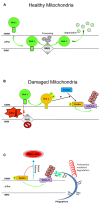
Cartoon illustrating steps in the mitochondrial clearance mediated by the Pink1–Parkin pathway. (A) In physiological conditions, Pink-1 is constitutively imported into healthy mitochondria via TIM/TOM complex to the inner membrane (IMM), cleaved by presenilin-associated rhomboid-like protease (PARL), and then proteolytically degraded. (B) Upon ΔΨ collapse, full-length Pink1 is not processed accumulating at the outer membrane (OMM) to recruit Parkin onto depolarized mitochondria. PINK1 autophosphorylation at Ser228 and Ser402 (P) is essential for efficient mitochondrial localization of Parkin. The PINK1-dependent Parkin phos-phorylation at Ser65, combined with unknown factor(s) (?), is required not only for its efficient translocation but also for the degradation of mito-chondrial proteins during mitophagy. (C) After being recruited on OMM, Parkin triggers mitophagy by ubiquitylating (Ub, K48, K63) several proteins including Mfns1/2, VDAC, TOM. Proteasome-mediated removal of Mfns1/2 not only inhibits mitochondrial fusion but also prevents its default rear-raggedright rangement into spheroids, allowing thus the damaged organelles to be recognized by the engulfing autophagosome. Cytosolic autophagy adaptor p62 (also known as sequestosome 1, SQSTM1) is also involved in mito-phagy as its K63-ubiquitin-binding domain (UBA) as well as an LC3- binding domain (LIR), recruits autophagosomes to ubiquitylated protein. For more information on Pink–Parkin-dependent mitophagy, please refer to recent excellent reviews (Twig and Shirihai, 2011; Vives-Bauza and Przedborski, 2011; Youle and Narendra, 2011; Ding and Yin, 2012; Jin and Youle, 2012). Freely adapted from Figure 6 of (Okatsu et al., 2012).
FIGURE 4
FIGURE 4
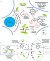
Cartoon illustrating the mitochondrial turnover which copes with the compartmentalized and distinct energetic requirements in the cell body, axon, and synaptic compartments of post-mitotic neurons. (A) In neurons, mitochondria travel long distances from the cell body out to distal dendritic and axonal terminals, where they subserve the ATP production and calcium homeostasis. The dynamic processes of biogenesis, fusion–fission regulate the mitochondrial function and quality control, by allowing them to adapt to spatial–temporal changes in cellular energy requirements. Selective autophagy begins with the nucleation of an isolation membrane (phagophore) which surrounds the damaged mitochondria to be degraded. RER could serve as membrane donors for autophagosome formation and the elongation of nascent double-membraned autophagic vesicle requires the coordinated assembly of Atg12–Atg5–Atg16L complex and LC3–PE conju-gates. (B) Newly formed autophagosomes move along microtubules in two directions – as a result of the opposing activities of the minus-end-directed motor protein dynein/dynactin and a plus-end-directed motor kinesin – and, finally, concentrate in perinuclear region (close to centrosome) where fuse with the lysosomes. Degradation of autophagosomal mitochondrial cargoes is then achieved by the acid hydrolases and the cathepsin proteases that are present in the lysosomal lumen.

References

    1. Allen G. F., Toth R., James J., Ganley I. G. (2013). Loss of iron triggers PINK1/Parkin-independent mitophagy. EMBO Rep. 14 1127–1135 10.1038/embor.2013.168 - DOI - PMC - PubMed
    1. Almeida A., Bolanos J., Moreno S. (2005). Cdh1/Hct1-APC is essential for the survival of postmitotic neurons. J. Neurosci. 25 8115–8121 10.1523/JNEUROSCI.1143-05.2005 - DOI - PMC - PubMed
    1. Amadoro G., Corsetti V., Atlante A., Florenzano F., Capsoni S., Bussani R., et al. (2012). Interaction between NH(2)-tau fragment and Aβ in Alzheimer’s disease mitochondria contributes to the synaptic deterioration. Neurobiol. Aging 33 833.e1–833.e25 10.1016/j.neurobiolaging.2011.08.001 - DOI - PubMed
    1. Amadoro G., Corsetti V., Florenzano F., Atlante A., Ciotti M. T., Mongiardi M. P., et al. (2014). AD-linked, toxic NH2 human tau increases the mitochondrial autophagy in neurons. Neurobiol. Dis. 62 489–507 10.1016/j.nbd.2013.10.018 - DOI - PubMed
    1. Amadoro G., Corsetti V., Stringaro A., Colone M., D’Aguanno S., Meli G., et al. (2010). A NH2 tau fragment targets neuronal mitochondria at AD synapses: possible implications for neurodegeneration. J. Alzheimers Dis. 21 445–470 10.3233/JAD-2010-100120 - DOI - PubMed

LinkOut - more resources