Review
. 2014 Feb 19:5:58.
doi: 10.3389/fphys.2014.00058. eCollection 2014.

The pannexins: past and present

Affiliations
Review

The pannexins: past and present

Stephen R Bond et al. Front Physiol. .

Abstract

The pannexins (Panxs) are a family of chordate proteins homologous to the invertebrate gap junction forming proteins named innexins. Three distinct Panx paralogs (Panx1, Panx2, and Panx3) are shared among the major vertebrate phyla, but they appear to have suppressed (or even lost) their ability to directly couple adjacent cells. Connecting the intracellular and extracellular compartments is now widely accepted as Panx's primary function, facilitating the passive movement of ions and small molecules along electrochemical gradients. The tissue distribution of the Panxs ranges from pervasive to very restricted, depending on the paralog, and are often cell type-specific and/or developmentally regulated within any given tissue. In recent years, Panxs have been implicated in an assortment of physiological and pathophysiological processes, particularly with respect to ATP signaling and inflammation, and they are now considered to be a major player in extracellular purinergic communication. The following is a comprehensive review of the Panx literature, exploring the historical events leading up to their discovery, outlining our current understanding of their biochemistry, and describing the importance of these proteins in health and disease.

Keywords: Panx1; Panx2; Panx3; biochemistry; distribution; gating; pannexin; structure.


Figures
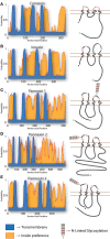
Figure 1
Figure 1
Topology of Cxs, Inxs, and Panxs. The predicted arrangement of these proteins within a membrane has been reported on multiple occasions (Hua et al., ; Panchin, ; Barbe et al., ; Magie and Martindale, 2008). To generate the predictions shown here, multiple pairwise alignments were calculated with the E-INS-i algorithm of MAFFT (Katoh and Standley, 2013) using (A) all 21 human Cxs, (B) 25 C. elegans Inxs, and (C–E) the three Chordate Panx isotypes using protein sequences from mouse, chicken, Xenopus, coelacanth, and Danio. Transmembrane (TM) regions and ER integration orientation were predicted with the OCTOPUS algorithm (Viklund and Elofsson, 2008), using the consensus sequences from each multiple alignment as input. All of the proteins have compelling support for 4 TM domains (blue), while the “inside-preference” (orange; based on clustering of positively charged and polar residues) assigned to the N-terminus and second loop strongly suggests that the termini are cytoplasmic. The topologies of each consensus sequence are shown to scale, with relative positions of extracellular loop cysteines and glycosylated asparagines indicated.
Figure 2
Figure 2
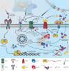
The role of Panx1 in induced innate immunity. When an invading microbe breaches an epithelial barrier, microbe-associated molecular patterns (MAMPs) are recognized by phagocytic cells [e.g., lipopolysaccharide (LPS) binding to toll-like receptors (TLR)], leading to an activation of the NF-κ B signaling cascade which up-regulates expression of the inflammasome component NLRP3, pro-IL-1β, and TNFα. Inflammasome assembly brings NLRP3 and pro-caspase1 together, using the adaptor protein ASC, and the whole complex associates with P2X7R. If extracellular ATP levels become elevated because of cellular distress in the vicinity (i.e., a “danger signal”), P2X7R activation allows calcium to flow into the cell and potassium to flow out-of the cell, along their respective concentration gradients. Lower intracellular potassium is correlated with inflammasome activation, leading to processing of pro-caspase1 into its active state. Panx1 is held in close proximity to P2X7R because of an interaction with residue P451 on the C-terminal end of the receptor, allowing local increases in extracellular potassium and intracellular calcium to stimulate channel opening, thus releasing more ATP into the extracellular space. This ATP further stimulates P2X receptors in the surrounding area, but also inhibits Panx1 channels as a negative feedback mechanism, preventing complete collapse of the electrochemical gradient. Meanwhile, liberated caspase1 will cleave the pro-peptide from IL-1β, which is then secreted from the cell along with TNFα. Receptors for both of these cytokines (e.g., IL-1R and TNFR) are also pro-inflammatory, and upon activation will initiate or perpetuate NF-κ B signaling. In addition, TNFR will induce the pro-apoptotic cascade that leads to caspase3 and caspase7 activation; both can clip the C-terminal tail of Panx1, which leads to a constitutively open channel. Several graphics included in this figure were derived from works deposited in the Wikimedia Commons (http://commons.wikimedia.org) by the following authors: LPS, by Mike Jones; DNA, by Sponk; background eukaryotic cell, by Mariana Ruiz; and mitochondria, by Kelvin Song.

References

    1. Abascal F., Zardoya R. (2012). LRRC8 proteins share a common ancestor with pannexins, and may form hexameric channels involved in cell-cell communication. Bioessays 37, 551–560 10.1002/bies.201100173 - DOI - PubMed
    1. Abascal F., Zardoya R. (2013). Evolutionary analyses of gap junction protein families. Biochim. Biophys. Acta 1828, 4–14 10.1016/j.bbamem.2012.02.007 - DOI - PubMed
    1. Adamczak S. E., de Rivero Vaccari J. P., Dale G., Brand Iii F. J., Nonner D., Bullock M., et al. (2014). Pyroptotic neuronal cell death mediated by the AIM2 inflammasome. J. Cereb. Blood Flow Metab. [Epub ahead of print]. 10.1038/jcbfm.2013.236 - DOI - PMC - PubMed
    1. Alberto A. V. P., Faria R. X., Couto C. G. C., Ferreira L. G. B., Souza C. A. M., Teixeira P. C. N., et al. (2013). Is pannexin the pore associated with the P2X7 receptor? Naunyn Schmiedebergs Arch. Pharmacol. 386, 775–787 10.1007/s00210-013-0868-x - DOI - PubMed
    1. Altun Z. F., Chen B., Wang Z. W., Hall D. H. (2009). High resolution map of Caenorhabditis elegans gap junction proteins. Dev. Dyn. 238, 1936–1950 10.1002/dvdy.22025 - DOI - PMC - PubMed