. 2014 Mar 6;9(6):e90773.
doi: 10.1371/journal.pone.0090773. eCollection 2014.

Motor cortex-evoked activity in reciprocal muscles is modulated by reward probability

Affiliations

Motor cortex-evoked activity in reciprocal muscles is modulated by reward probability

Makoto Suzuki et al. PLoS One. .

Abstract

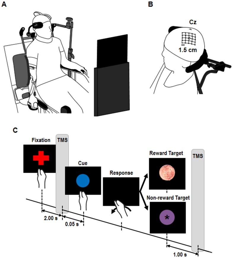
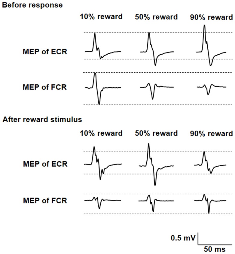
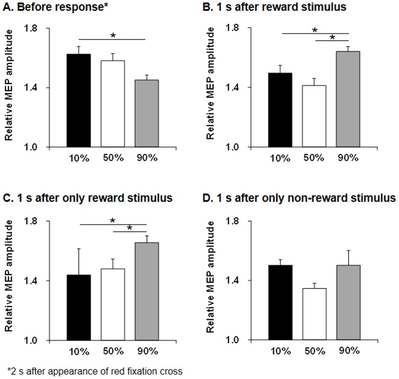
Horizontal intracortical projections for agonist and antagonist muscles exist in the primary motor cortex (M1), and reward may induce a reinforcement of transmission efficiency of intracortical circuits. We investigated reward-induced change in M1 excitability for agonist and antagonist muscles. Participants were 8 healthy volunteers. Probabilistic reward tasks comprised 3 conditions of 30 trials each: 30 trials contained 10% reward, 30 trials contained 50% reward, and 30 trials contained 90% reward. Each trial began with a cue (red fixation cross), followed by blue circle for 1 s. The subjects were instructed to perform wrist flexion and press a button with the dorsal aspect of middle finger phalanx as quickly as possible in response to disappearance of the blue circle without looking at their hand or the button. Two seconds after the button press, reward/non-reward stimulus was randomly presented for 2-s duration. The reward stimulus was a picture of Japanese 10-yen coin, and each subject received monetary reward at the end of experiment. Subjects were not informed of the reward probabilities. We delivered transcranial magnetic stimulation of the left M1 at the midpoint between center of gravities of agonist flexor carpi radialis (FCR) and antagonist extensor carpi radialis (ECR) muscles at 2 s after the red fixation cross and 1 s after the reward/non-reward stimuli. Relative motor evoked potential (MEP) amplitudes at 2 s after the red fixation cross were significantly higher for 10% reward probability than for 90% reward probability, whereas relative MEP amplitudes at 1 s after reward/non-reward stimuli were significantly higher for 90% reward probability than for 10% and 50% reward probabilities. These results implied that reward could affect the horizontal intracortical projections in M1 for agonist and antagonist muscles, and M1 excitability including the reward-related circuit before and after reward stimulus could be differently altered by reward probability.


Conflict of interest statement

Competing Interests: The authors have declared that no competing interests exist.

Figures

Figure 1
Figure 1. Experimental setup.
(A) Change in primary motor cortex (M1) excitability for agonist and antagonist muscles during probabilistic reward tasks was investigated. Subjects were seated comfortably in a chair. The right arm hung to the side in a relaxed posture, with the palm and forearm placed on the equipment. (B) Schematic of a head with a grid showing the stimulated scalp sites. Cz represents the intersection of nasion-inion and the interaural lines. (C) Experimental design in probabilistic reward task. Probabilistic reward tasks comprised 3 conditions of 30 trials: 30 trials contained 10% reward stimulus and the remaining trials contained a non-target stimulus, 30 trials contained 50% reward stimulus, and 30 trials contained 90% reward stimulus. The inter-trial interval was randomized between 7–8 s. Single-pulse transcranial magnetic stimulation (TMS) was delivered at 2 s after appearance of the red fixation cross and 1 s after appearance of the reward/non-reward stimuli.
Figure 2
Figure 2. Two-dimensional maps.
The color code of each map of FCR (A) and ECR (B) muscles ranges from gray (0 mV) to white (0.5 mV or over). The map areas of the FCR and ECR muscles clearly overlapped, although they were spread differently. The center of gravity (black circle) of the FCR muscle was located at x (anteroposterior)  = 6.5±2.6 mm and y (mediolateral)  = 56.5±2.3 mm and that of the ECR muscle was located at x = 4.5±3.6 mm and y = 56.4±2.7 mm. FCR: flexor carpi radialis; ECR: extensor carpi radialis.
Figure 3
Figure 3. Electromyography traces of the right FCR and ECR muscles in one representative subject.
MEP amplitude of the FCR muscle at 2 s before response was the highest for 10% reward probability during the task, whereas that of the ECR muscle was the lowest for 10% reward probability. However, MEP amplitude of the FCR muscle at 1 s before response was the highest for 90% reward probability during the task, whereas that of the ECR muscle was the lowest for 90% reward probability. MEP, motor-evoked potential; FCR, flexor carpi radialis; ECR, extensor carpi radialis.
Figure 4
Figure 4. Bar graphs of relative MEP amplitudes for FCR and ECR muscles.
Relative MEP amplitude at 2(A) and at 1 s after reward/non-reward stimuli (B) during the task. Relative MEP amplitude at 2 s after the red fixation cross was significantly higher for 10% reward probability than for 90% reward probability (p = 0.008) during the task, whereas relative MEP amplitude at 1 s after reward/non-reward stimuli was significantly higher for 90% reward probability than for 10% (p = 0.001) and 50% (p = 0.001) reward probabilities. Bar graphs of relative MEP amplitudes for FCR and ECR muscles at 1 s after only reward stimuli presentation (C) and only non-reward stimuli presentation (D) during the task. Relative MEP amplitude at 1 s after only reward stimuli presentation was significantly higher for 90% reward probability than for 10% (p<0.0001) and 50% (p = 0.006) reward probabilities. However, relative MEP amplitudes for FCR and ECR muscles at 1 s after only non-reward stimuli presentation were not significantly changed. MEP, motor-evoked potential; FCR, flexor carpi radialis; ECR, extensor carpi radialis.
Figure 5
Figure 5. Bar graphs of RMT, SICI, and SAI before and after probabilistic reward tasks.
RMT of FCR (A) and ECR (B) for 10% reward probability, RMT of FCR (C) and ECR (D) for 50% reward probability, RMT of FCR (E) and ECR (F) for 90% reward probability, SICI of FCR (G) and ECR (H) for 10% reward probability, SICI of FCR (I) and ECR (J) for 50% reward probability, SICI of FCR (K) and ECR (L) for 90% reward probability, SAI of FCR (M) and ECR (N) for 10% reward probability, SAI of FCR (O) and ECR (P) for 50% reward probability, and SAI of FCR (Q) and ECR (R) for 90% reward probability. Only the SICI of the FCR was significantly decreased after 10% probabilistic reward tasks (p = 0.0008). RMT, resting motor threshold; SICI, short-interval intracortical inhibition; SAI, short-latency afferent inhibition; FCR, flexor carpi radialis; ECR, extensor carpi radialis.

References

    1. Wickens JR, Reynolds JN, Hyland BI (2003) Neural mechanisms of reward-related motor learning. Curr Opin Neurobiol 13: 685–690. - PubMed
    1. Centonze D, Grande C, Saulle E, Martin AB, Gubellini P, et al. (2003) Distinct roles of D1 and D5 dopamine receptors in motor activity and striatal synaptic plasticity. J Neurosci 23: 8506–8512. - PMC - PubMed
    1. Calabresi P, Picconi B, Tozzi A, Di Filippo M (2007) Dopamine-mediated regulation of corticostriatal synaptic plasticity. Trends Neurosci 30: 211–219. - PubMed
    1. Davis EJ, Coyne C, McNeill TH (2007) Intrastriatal dopamine D1 antagonism dampens neural plasticity in response to motor cortex lesion. Neuroscience 146: 784–791. - PMC - PubMed
    1. Lang N, Speck S, Harms J, Rothkegel H, Paulus W, et al. (2008) Dopaminergic potentiation of rTMS-induced motor cortex inhibition. Biol Psychiatry 63: 231–233. - PubMed

Publication types

LinkOut - more resources