. 2014 Mar 6;9(3):e89375.
doi: 10.1371/journal.pone.0089375. eCollection 2014.

Characterisation of leukocytes in a human skin blister model of acute inflammation and resolution

Affiliations

Characterisation of leukocytes in a human skin blister model of acute inflammation and resolution

William Jenner et al. PLoS One. .

Abstract

There is an increasing need to understand the leukocytes and soluble mediators that drive acute inflammation and bring about its resolution in humans. We therefore carried out an extensive characterisation of the cantharidin skin blister model in healthy male volunteers. A novel fluorescence staining protocol was designed and implemented, which facilitated the identification of cell populations by flow cytometry. We observed that at the onset phase, 24 h after blister formation, the predominant cells were CD16hi/CD66b+ PMNs followed by HLA-DR+/CD14+ monocytes/macrophages, CD11c+ and CD141+ dendritic cells as well as Siglec-8+ eosinophils. CD3+ T cells, CD19+ B cells and CD56+ NK cells were also present, but in comparatively fewer numbers. During resolution, 72 h following blister induction, numbers of PMNs declined whilst the numbers of monocyte/macrophages remain unchanged, though they upregulated expression of CD16 and CD163. In contrast, the overall numbers of dendritic cells and Siglec-8+ eosinophils increased. Post hoc analysis of these data revealed that of the inflammatory cytokines measured, TNF-α but not IL-1β or IL-8 correlated with increased PMN numbers at the onset. Volunteers with the greatest PMN infiltration at onset displayed the fastest clearance rates for these cells at resolution. Collectively, these data provide insight into the cells that occupy acute resolving blister in humans, the soluble mediators that may control their influx as well as the phenotype of mononuclear phagocytes that predominate the resolution phase. Further use of this model will improve our understanding of the evolution and resolution of inflammation in humans, how defects in these over-lapping pathways may contribute to the variability in disease longevity/chronicity, and lends itself to the screen of putative anti-inflammatory or pro-resolution therapies.


Conflict of interest statement

Competing Interests: The authors have declared that no competing interests exist.

Figures

Figure 1
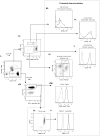
Figure 1. Characterisation of peripheral blood leukocytes from healthy volunteers- I.
Representative dot plots for flow cytometric gating are shown for healthy male volunteers (n = 17). Blood was drawn from the forearm not bearing skin blisters. Erythrocytes were lysed and the remaining leukocytes incubated with antibodies and processed by flow cytometry. Gating strategies firstly identified CD3+ T cells, CD19+ B cells and CD56+ CD16+/− NK cells. The remaining lymphocyte-deplete population was gated into HLA-DR+ and HLA-DR cells. Arrows indicate gating strategy.
Figure 2
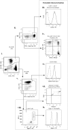
Figure 2. Characterisation of peripheral blood leukocytes from healthy volunteers- II.
HLA-DR+ and HLA-DR cells identified in Figure 1 were further analysed. HLA-DR+ cells were characterised into CD14hi/CD16 , CD14hi/CD16+, CD14lo/CD16+ monocytes and CD14/CD16 dendritic cells. HLA-DR cells comprised of typical PMNs (CD16hi, CD66+) and a CD16lo population. On extended characterisation, HLA-DR/CD16lo cells were identified as Siglec-8+ eosinophils and the HLA-DR+/CD14/CD16 dendritic cells comprised of CD141+ and CD11c+ dendritic cell subpopulations. Arrows indicate gating strategy.
Figure 3
Figure 3. Characterisation of inflammatory cell infiltrates into skin blisters at 24(onset) – I.
Representative dot plots for flow cytometric gating are shown for healthy male volunteers (n = 17). Blister contents were collected from one blister 24 h after application of cantharidin in 3% sodium citrate, with cells separated from oedema by centrifugation. Leukocytes were enumerated by haemocytometer and oedema volume recorded. Leukocytes were incubated with antibodies and processed by flow cytometry. Gating strategies firstly identified CD3+ T cells, CD19+ B cells and CD56+CD16+/− NK cells. The remaining lymphocyte-deplete population was gated into HLA-DR+ and HLA-DR cells. Arrows indicate gating strategy.
Figure 4
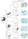
Figure 4. Characterisation of inflammatory cell infiltrates into skin blisters at 24(onset) – II.
HLA-DR+ and HLA-DR cells identified in Figure 3 were further analysed. HLA-DR+ cells were characterised into CD14+/CD16lo monocytes/macrophages and HLA-DR+/CD14/CD16 dendritic cells. HLA-DR cells comprised of typical PMNs (CD16hi, CD66+) and a CD16lo population. On extended characterisation, HLA-DR/CD16lo were identified as a mixture of Siglec-8+ eosinophils and Annexin V/7AAD+ apoptotic/dead PMNs. The HLA-DR+/CD14/CD16 dendritic cells comprised of CD141+ and CD11c+ dendritic cell subpopulations. Arrows indicate gating strategy.
Figure 5
Figure 5. Characterisation of inflammatory cell infiltrates into skin blisters at 72(resolution) – I.
Representative dot plots for flow cytometric gating are shown for healthy male volunteers (n = 17). Blister contents were collected from the remaining blister 72 h after application of cantharidin in 3% sodium citrate, with cells separated from oedema by centrifugation. Leukocytes were enumerated by haemocytometer and oedema volume recorded. Leukocytes were incubated with antibodies and processed by flow cytometry. Gating strategies firstly identified CD3+ T cells, CD19+ B cells and CD56+CD16+/− NK cells. The remaining lymphocyte-deplete population was gated into HLA-DR+ and HLA-DR cells. Arrows indicate gating strategy.
Figure 6
Figure 6. Characterisation of inflammatory cell infiltrates into skin blisters at 72(resolution) – II.
HLA-DR+ and HLA-DR cells identified in Figure 5 were further analysed. HLA-DR+ cells were characterised into CD14+/CD16hi monocytes/macrophages and CD14 CD16 dendritic cells. HLA-DR cells comprised of typical PMNs (CD16hi, CD66b+) and CD16lo population. On extended characterisation, HLA-DR/CD16lo were identified as Siglec-8+ eosinophils and the HLA-DR+/CD14/CD16 dendritic cells comprised of CD141+ and CD11c+ dendritic cell subpopulations. Arrows indicate gating strategy.
Figure 7
Figure 7. Total leukocyte and oedema profile in resolving skin blisters in humans.
Blister content was collected in 3% sodium citrate 24 h and 72 h after application of cantharidin with cells separated from oedema by centrifugation. Red blood cells were lysed and the remaining leukocytes counted by haemocytometer while oedema volume recorded. Temporal differences from 24 to 72 h for total cells, leukocyte concentration and oedema volume are shown (n = 17).
Figure 8
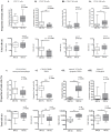
Figure 8. Leukocyte subtype profiles in resolving skin blisters in humans.
Blister content was collected in 3% sodium citrate 24 h and 72 h after application of cantharidin with cells separated from oedema by centrifugation. Red blood cells were lysed and the remaining leukocytes counted by haemocytometer. Leukocytes were incubated with fluorescent antibodies, processed for flow cytometry, and gated according to Figures 2 and 3. Temporal changes in cell populations from 24 h to 72 h are shown (n = 17).
Figure 9
Figure 9. Cytokine profiles in resolving skin blisters in humans.
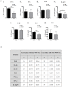
Blister content was collected in 3% sodium citrate 24 h and 72 h after application of cantharidin with cells separated from oedema by centrifugation. Red blood cells were lysed and the remaining leukocytes counted by haemocytometer while oedema volume recorded and processed by assay for cytokine concentration (A). Correlations were made between cytokine concentration and PMN infiltration at 24 h and 72 h (B) (n = 17).
Figure 10
Figure 10. Lipid mediators in resolving skin blisters in humans.
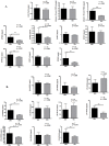
Blister content was collected in 3% sodium citrate 24 h and 72 h after application of cantharidin with cells separated from oedema by centrifugation. Red blood cells were lysed and the remaining leukocytes counted by haemocytometer while oedema volume recorded and analysed by liquid chromatography coupled to electrospray ionization tandem mass spectrometry for eicosanoid levels (n = 17).
Figure 11
Figure 11. Correlations in cell profiles from onset to resolution.
A wide range of PMN numbers infiltrate skin blisters early in the response (onset), and an immune response induces appropriate resolution. Correlation between change in PMN numbers (72 h minus 24 h) and total PMN numbers at onset are expressed as either total cells or as percentages.

References

    1. Lawrence T, Willoughby DA, Gilroy DW (2002) Anti-inflammatory lipid mediators and insights into the resolution of inflammation. Nature reviews Immunology 2: 787–795. - PubMed
    1. Vane JR, Botting RM (1995) New insights into the mode of action of anti-inflammatory drugs. Inflammation research : official journal of the European Histamine Research Society [et al] 44: 1–10. - PubMed
    1. Serhan CN, Savill J (2005) Resolution of inflammation: the beginning programs the end. Nature immunology 6: 1191–1197. - PubMed
    1. Colville-Nash P, Lawrence T (2003) Air-pouch models of inflammation and modifications for the study of granuloma-mediated cartilage degradation. Methods in molecular biology 225: 181–189. - PubMed
    1. Gabor M (2003) Models of acute inflammation in the ear. Methods in molecular biology 225: 129–137. - PubMed

Publication types

MeSH terms