. 2014 Mar 6;10(3):e1004178.
doi: 10.1371/journal.pgen.1004178. eCollection 2014 Mar.

Nucleosome acidic patch promotes RNF168- and RING1B/BMI1-dependent H2AX and H2A ubiquitination and DNA damage signaling

Affiliations

Nucleosome acidic patch promotes RNF168- and RING1B/BMI1-dependent H2AX and H2A ubiquitination and DNA damage signaling

Justin W Leung et al. PLoS Genet. .

Abstract

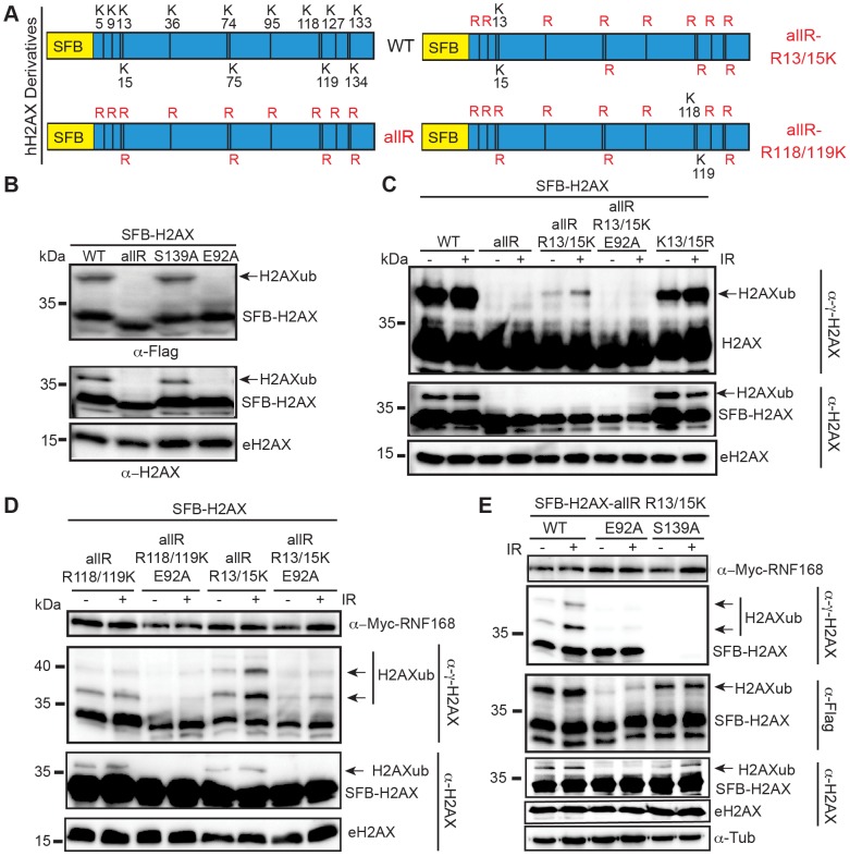
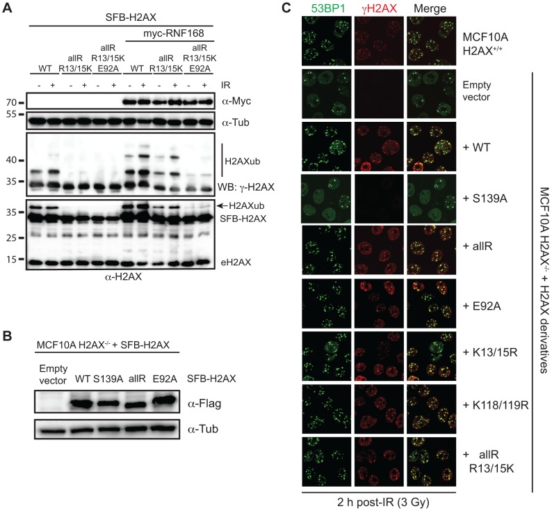
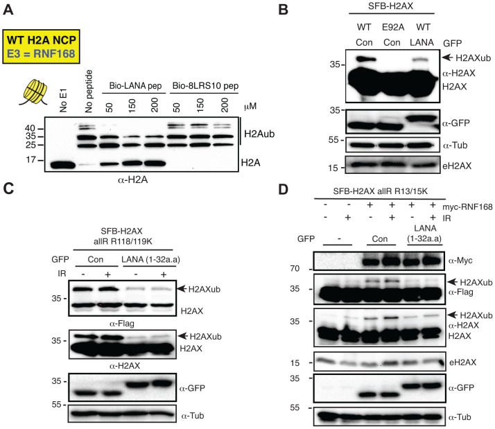
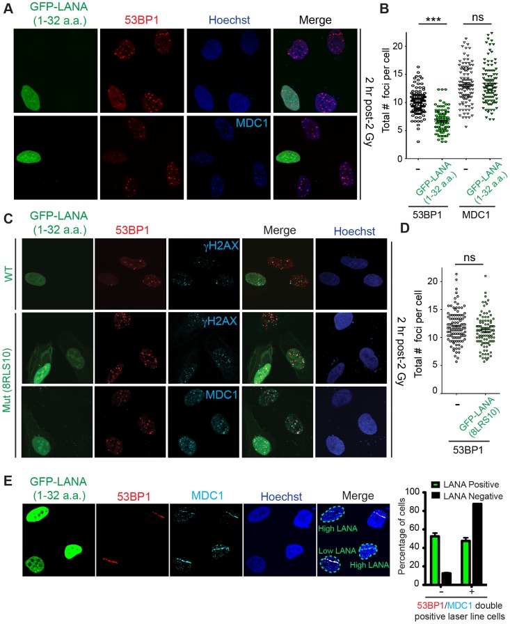
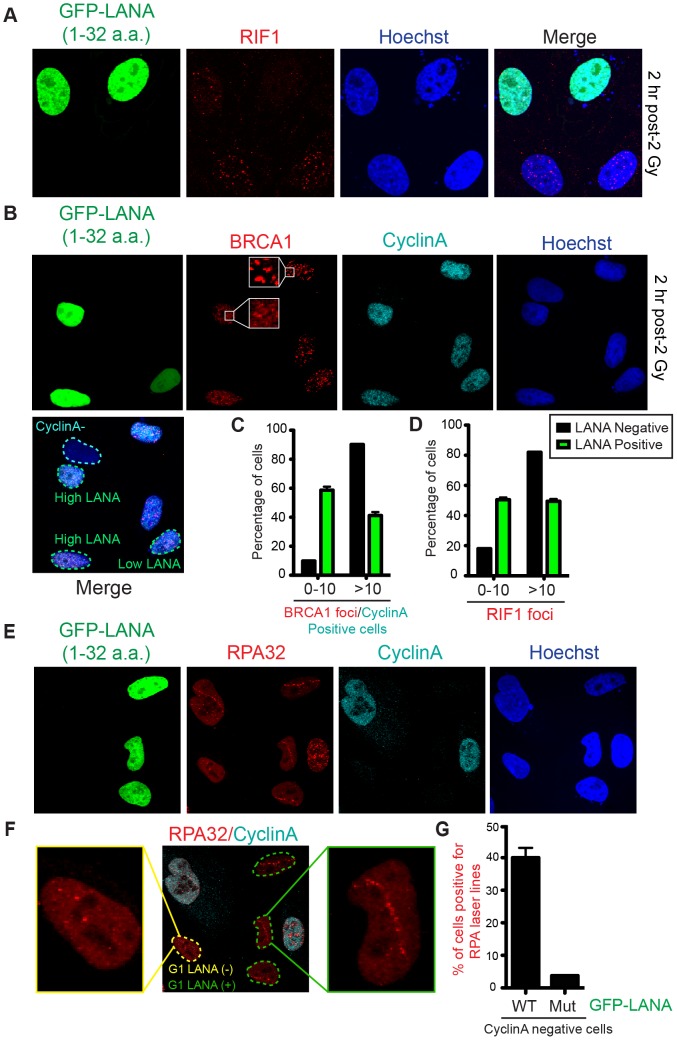
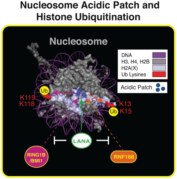
Histone ubiquitinations are critical for the activation of the DNA damage response (DDR). In particular, RNF168 and RING1B/BMI1 function in the DDR by ubiquitinating H2A/H2AX on Lys-13/15 and Lys-118/119, respectively. However, it remains to be defined how the ubiquitin pathway engages chromatin to provide regulation of ubiquitin targeting of specific histone residues. Here we identify the nucleosome acid patch as a critical chromatin mediator of H2A/H2AX ubiquitination (ub). The acidic patch is required for RNF168- and RING1B/BMI1-dependent H2A/H2AXub in vivo. The acidic patch functions within the nucleosome as nucleosomes containing a mutated acidic patch exhibit defective H2A/H2AXub by RNF168 and RING1B/BMI1 in vitro. Furthermore, direct perturbation of the nucleosome acidic patch in vivo by the expression of an engineered acidic patch interacting viral peptide, LANA, results in defective H2AXub and RNF168-dependent DNA damage responses including 53BP1 and BRCA1 recruitment to DNA damage. The acidic patch therefore is a critical nucleosome feature that may serve as a scaffold to integrate multiple ubiquitin signals on chromatin to compose selective ubiquitinations on histones for DNA damage signaling.


Conflict of interest statement

The authors have declared that no competing interests exist.

Figures

Figure 1
Figure 1. Mutation of the acidic patch impairs human H2AX and H2A ubiquitination.
(A) Schematic of all H2AX lysines (K) and mutant derivatives. allR represents an all lysine (K) to arginine (R) version of H2AX. Additional site-specific reversions from arginine to lysine within the allR H2AX derivate are indicated. (B) H2AX-allR and acidic patch mutation E92A reduces H2AXub. WT or E92A H2AX/H2A constructs were transfected into HEK293T cells and analyzed by western blotting with the indicated antibodies. Arrows indicate ub forms. (SFB = S-tag, Flag epitope tag, and streptavidin-binding peptide tag; e = endogenous). Molecular weights (kDa) are indicated on the left of each panel. HEK293T cells were used for all cellular assays. (C) H2AX-K13/15 dependent ubiquitination requires the acidic patch. H2AX and derivatives were expressed in HEK293T cells (−) or (+) ionizing radiation (IR, 20 Gy). Samples were analyzed as in A 6 h post-IR treatment. (D) H2AX-K13/15 and K118/119-dependent ubiquitination requires the acidic patch. Cells were co-transfected with H2AX and derivatives along with Myc-RNF168 and analyzed as in C. (E) Phospho-competent H2AX S139 is not required in cis for H2AX K13/15ub. Cells were analyzed as in C. tub = tubulin loading control.
Figure 2
Figure 2. RING1B/BMI1- and RNF168-dependent ubiquitination of H2AX/H2A requires the nucleosome acidic patch in vitro.
(A) Schematic for in vitro reconstitution of nucleosome core particles (NCPs). (B) Bacterially expressed and purified human histones. Histones were expressed, purified and reconstituted as described in methods. (C) Analysis of in vitro reconstituted NCPs. The 147 bp 601 DNA fragment was analyzed alone or after NCP reconstitution. DNA ladder indicates size (bp). (D and E) RING1B/BMI1 and RNF168 readily ubiquitinate H2AX within WT NCPs but not NCPs containing a mutation in the acidic patch (H2AX-E92A). In vitro Ub assays (4 h) were performed as described in methods. (F) RNF168 ubiquitinates WT H2AX and H2AX-E92A similarly when assayed in the context of free histones. Assays were performed as in E except with free histones and reactions were performed overnight. (G and H) RING1B/BMI1 and RNF168 readily ubiquitinate H2A within WT NCPs but not NCPs containing a mutation in the acidic patch (H2A-E92A). Experiments were performed as in D and E using H2A. (I) RNF168 ubiquitinates free WT H2A and H2A-E92A similarly. Experiments performed as in F.
Figure 3
Figure 3. Regulation of H2AX-K13/K15ub by RNF168 requires the acidic patch.
(A) Maximum H2AX-K13/15ub levels (−) or (+) RNF168 is dependent on the acidic patch. HEK293T cells were transfected with H2AX and derivatives either (−) or (+) RNF168 and analyzed as in Figure 1D. (B) Human H2AX and derivatives reconstituted in MCF10A H2AX−/− cells. Western blot analysis of the indicated MCF10A H2AX−/− stable cell lines. (C) H2AXub is dispensable for 53BP1 foci formation after DNA damage. Reconstituted MCF10A H2AX−/− cells stably expressing H2AX and H2AX mutants were analyzed by immunofluorescence (IF) with the indicated antibodies. Cells were treated with 3 Gy IR and analyzed by IF 2 h post-IR.
Figure 4
Figure 4. The KSHV LANA peptide inhibits histone ubiquitination in vitro and in vivo.
(A) The acidic patch interaction region of LANA inhibits RNF168-dependent H2Aub in vitro. In vitro Ub assays were performed (−) or (+) either LANA peptide or a mutant LANA peptide (8LRS10) that does not interact with the nucleosome acidic patch. Assays were performed as in Figure 2 with increasing concentrations of peptides (µM) as indicated (4 h reactions). (B) Expression of GFP-LANA (1–32a.a.) reduces H2AXub. HEK293T cells were transfected with the indicated constructs and analyzed by western blotting as in Figure 1B. (C) GFP-LANA (1–32a.a.) reduces H2AXub at K118/119. Experiments were performed as in B using H2AX-allR-R118/119K with or without IR treatment. (D) Expression of GFP-LANA (1–32a.a.) reduces RNF168-dependent H2AX-K13/15ub. Experiments were performed and analyzed as in Figure 3A with the indicated constructs, with or without IR. Arrows indicate H2AXub protein species. e = endogenous; con = control GFP alone.
Figure 5
Figure 5. The nucleosome acidic patch is required in vivo for the DDR in human cells.
(A and B) In vivo expression of the acidic patch interacting portion of LANA (1–32 amino acids) reduces 53BP1, but not MDC1, IRIF (ionizing radiation induced-foci). Human U2OS cells were transfected with GFP-LANA (1–32a.a.) followed by 2 Gy IR-treatment. Cells were analyzed by IF with the indicated antibodies 2 h post-IR. Representative IF images are shown. Nuclear DNA was visualized by Hoechst 33342 staining. Quantification of A is shown in B. 53BP1 and MDC1 IRIF were counted and graphed for cells (−) or (+) GFP-LANA (1–32a.a.). N = 3, >100 cells analyzed/experiment, error bars = SEM. Student's t-tests (paired) were performed and results indicated. *** = p-value<0.001, ns = not significant (i.e. p-value>0.05). (C) IF analysis of DDR factor foci formation after IR treatment in GFP-LANA and mutant GFP-LANA-8LRS10 expressing cells. Cells were treated with 2 Gy IR and processed for IF 2 h post-IR. IF analysis was performed as in A. (D) Quantification of 53BP1 IRIF from C. Graph represents values obtained from two independent experiments where foci from >100 cells were scored for GFP-LANA-8LRS10 expressing cells and non-GFP expressing cells. Error bars = SEM. Statistical analysis was performed as in B. (E) GFP-LANA (1–32a.a.) impairs recruitment of 53BP1 to laser damage. U2OS cells were transfected with GFP-LANA (1–32a.a.) followed by laser micro-irradiation. Cells were fixed and stained with antibodies as indicated 2 h post-laser damage. Quantification of 53BP1 and MDC1 laser lines were obtained from >50 damaged cells from two independent experiments. Error bars = SEM.
Figure 6
Figure 6. Nucleosome acidic patch promotes RNF168-dependent DDR signaling and inhibition of DNA resection in G1.
(A and B) RIF1 and BRCA1 IRIF are impaired in GFP-LANA (1–32a.a.) expressing cells. Experiments were performed as in Figure 5A. Representative images are shown. CyclinA negative and high/low LANA expressing cells are indicated in the merged image. Note: CyclinA marks S/G2 cells. (C and D) Quantification of RIF1 and BRCA1/CyclinA-positive IRIF from A and B. Graphs represent values obtained from two independent experiments where foci from >100 cells were quantified. Error bars = SEM. (E) GFP-LANA (1–32a.a.) expressing cells exhibit DNA end-resection as detected by RPA accumulation at laser damage in G1 (CyclinA-negative) cells. Experiments were performed as in Figure 5E with indicated antibodies after 4 h post micro-irradiation. (F) RPA32 laser lines in CyclinA-negative cells without or with GFP-LANA (1–32a.a.) expression are indicated by yellow and green dotted lines respectively. Enlarged images from each category are shown. All cells have been laser damaged. (G) Quantification of F and G from either WT GFP-LANA (1–32a.a.) or mutant (Mut) GFP-LANA-8LRS10 expressing cells. Laser damaged CylclinA negative cells positive for LANA expression were scored for RPA laser line formation. Graph represents data obtained from >50 cells from two independent experiments. Error bars = SEM.
Figure 7
Figure 7. The nucleosome acidic patch and histone ubiquitination.
Summary of our results within the context of the nucleosome structure. The acidic patch is required for RNF168- and RING1B/BMI1-dependent histone ubiquitination and LANA inhibits these processes. See text for details. Nucleosome structure was created in Pymol as previously described .

References

    1. Kouzarides T (2007) Chromatin modifications and their function. Cell 128: 693–705. - PubMed
    1. Miller KM, Jackson SP (2012) Histone marks: repairing DNA breaks within the context of chromatin. Biochem Soc Trans 40: 370–376. - PubMed
    1. Rogakou EP, Pilch DR, Orr AH, Ivanova VS, Bonner WM (1998) DNA double-stranded breaks induce histone H2AX phosphorylation on serine 139. J Biol Chem 273: 5858–5868. - PubMed
    1. Paull TT, Rogakou EP, Yamazaki V, Kirchgessner CU, Gellert M, et al. (2000) A critical role for histone H2AX in recruitment of repair factors to nuclear foci after DNA damage. Curr Biol 10: 886–895. - PubMed
    1. Rogakou EP, Boon C, Redon C, Bonner WM (1999) Megabase chromatin domains involved in DNA double-strand breaks in vivo. J Cell Biol 146: 905–916. - PMC - PubMed

Publication types