. 2014 Mar 6;9(3):e90527.
doi: 10.1371/journal.pone.0090527. eCollection 2014.

Clinical significance of markers of collagen metabolism in rheumatic mitral valve disease

Affiliations

Clinical significance of markers of collagen metabolism in rheumatic mitral valve disease

Tanima Banerjee et al. PLoS One. .

Abstract

Background: Rheumatic Heart Disease (RHD), a chronic acquired heart disorder results from Acute Rheumatic Fever. It is a major public health concern in developing countries. In RHD, mostly the valves get affected. The present study investigated whether extracellular matrix remodelling in rheumatic valve leads to altered levels of collagen metabolism markers and if such markers can be clinically used to diagnose or monitor disease progression.

Methodology: This is a case control study comprising 118 subjects. It included 77 cases and 41 healthy controls. Cases were classified into two groups- Mitral Stenosis (MS) and Mitral Regurgitation (MR). Carboxy-terminal propeptide of type I procollagen (PICP), amino-terminal propeptide of type III procollagen (PIIINP), total Matrix Metalloproteinase-1(MMP-1) and Tissue Inhibitor of Metalloproteinase-1 (TIMP-1) were assessed. Histopathology studies were performed on excised mitral valve leaflets. A p value <0.05 was considered statistically significant.

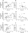
Results: Plasma PICP and PIIINP concentrations increased significantly (p<0.01) in MS and MR subjects compared to controls but decreased gradually over a one year period post mitral valve replacement (p<0.05). In MS, PICP level and MMP-1/TIMP-1 ratio strongly correlated with mitral valve area (r = -0.40; r = 0.49 respectively) and pulmonary artery systolic pressure (r = 0.49; r = -0.49 respectively); while in MR they correlated with left ventricular internal diastolic (r = 0.68; r = -0.48 respectively) and systolic diameters (r = 0.65; r = -0.55 respectively). Receiver operating characteristic curve analysis established PICP as a better marker (AUC = 0.95; 95% CI = 0.91-0.99; p<0.0001). A cut-off >459 ng/mL for PICP provided 91% sensitivity, 90% specificity and a likelihood ratio of 9 in diagnosing RHD. Histopathology analysis revealed inflammation, scarring, neovascularisation and extensive leaflet fibrosis in diseased mitral valve.

Conclusions: Levels of collagen metabolism markers correlated with echocardiographic parameters for RHD diagnosis.


Conflict of interest statement

Competing Interests: The authors have declared that no competing interests exist.

Figures

Figure 1
Figure 1. Plasma concentrations of circulating biomarkers of collagen turnover in normal, MS and MR subjects.
(A) Mean plasma PICP in control, MS and MR subjects before (Pre Op) valve replacement surgery. (B) Progressive reduction in plasma PICP concentration one month and one year or above following mitral valve replacement. (C) Mean plasma PIIINP in control, MS and MR subjects before (Pre Op) valve replacement surgery. (D) Progressive decrease in plasma PIIINP concentration one month and one year or more after mitral valve replacement. (E) Mean plasma concentration of total MMP-1 in control, MS and MR subjects. (F) Mean TIMP-1 concentration in control, MS and MR subjects. (G) Plasma MMP-1/TIMP-1 ratio in control, MS and MR subjects.
Figure 2
Figure 2. Receiver operating characteristics curve for biomarkers in rheumatic heart disease subjects.
Figure 3
Figure 3. Relationship between plasma markers of collagen metabolism and severity of rheumatic mitral stenosis.
(A) Inverse correlations of plasma PICP (y = −17.241x+2654.1; p = 0.01) and PIIINP (y = −4.6576x+938.36; p = 0.15) concentration with mitral valve area(MVA). (B) Direct correlation (y = 0.0127x−0.582; p = 0.03) between plasma MMP-1/TIMP-1 ratio and MVA. (C) Direct correlation of plasma PICP (y = 24.155x+186.83;p = 0.02) and almost no correlation of plasma PIIINP (y = −0.4083+634.78;p = 0.91) with pulmonary artery systolic pressure (PASP). (D) Inverse correlation (y = −0.0091x+0.8791; p = 0.05) between plasma MMP-1/TIMP-1 ratio and PASP.
Figure 4
Figure 4. Relationship between plasma PICP levels and hemodynamic parameters in rheumatic mitral regurgitation.
(A) Direct correlation of plasma PICP (y = 28.83x−875.54;p = 0.0001) and no correlation of plasma PIIINP (y = 1.5469x+471.72; p = 0.79) with left ventricular diameter at diastole (LVIDd). (B) Inverse correlation (y = −0.0181x+1.6878; p = 0.04) between plasma MMP-1/TIMP-1 ratio and ventricular diameter at diastole (LVIDd). (C) Direct correlation of plasma PICP (y = 34.933x−506.05; p = 0.0002) and no correlation of plasma PIIINP (y = −3.3609x+697.11; p = 0.64) with left ventricular internal diameter at systole (LVIDs). (D) Inverse correlation(y = −0.0241x+1.523;p = 0.01) between plasma MMP-1/TIMP-1 ratioand LVIDs. (E) Direct correlation of plasma PICP (y = 3620.7x+53.656; p<0.0001) and weak correlation of plasma PIIINP (y = 593.3x+430.94; p = 0.39) with left ventricular mass (LVM). (F) Inverse correlation (y = −1.9248x+1.0243;p = 0.03) between plasma MMP-1/TIMP-1 ratio and LVM.
Figure 5
Figure 5. Histopathology of heart valve.
(A) Representative images (100× magnification) of hematoxylin-eosin stained sections of 1 normal heart valve (control) and 3 rheumatic valve samples (RHD). Rheumatic mitral valve tissue section shows abundance of inflammatory cells (arrow head), fibrosis (blue arrow) and neovascularisation (black arrow). Normal mitral valve section shows wavy arrangement of collagen fibres (black arrow). Scale bar represents 50 µm. (B) Representative images (100×) of Masson's trichrome stained cross sections showing dense collagen deposition (arrow) in mitral valve tissue of RHD patient compared to loose parallel pattern of collagen (arrow) in normal heart valve. Scale bar represents 50 µM.
Figure 6
Figure 6. Assessment of collagen deposition by picrosirius red staining and immunostaining methods.
(A) Representative images (20× magnification) of picrosirius red stained sections of 1 normal heart valve (control) and 3 rheumatic mitral valve samples (RHD). Stained sections were observed using a binocular polarized light microscope. Under polarized light, birefringence is specific for collagen where red colour shows fibrillar type I collagen and yellow green colour indicates reticular type III collagen. Arrow indicates scattered deposition of collagen type I in diseased valve Scale bar represents 100 µm. (B) Total collagen intensity in control vs. RHD mitral valve tissue sections. (C) Type I collagen mean intensity in control vs. RHD mitral valve cross sections. (D) Type III collagen mean intensity in control vs. RHD mitral valve cross sections. (E) Ratio of Type I to Type III collagen in control vs. RHD mitral valve sections.(F) Representative images of immuno stained sections of 1 normal heart valve (control) and 3 rheumatic mitral valve samples (RHD) showing (arrow marked) collagen type 1 deposition. Scale bar represents 45 µm. *p<0.05 vs. control,***p<0.0001 vs. control. Here “n” denotes total number of tissue sections.

References

    1. Marijon E, Mirabel M, Celermajer DS, Jouven X (2012) Rheumatic Heart Disease. Lancet 379: 953–964. - PubMed
    1. GuilhermeL, Köhler KF, Postol E, Kalil J (2011) Genes, autoimmunity and pathogenesis of rheumatic heart disease. Ann Pediatr Cardiol 4: 13–21. - PMC - PubMed
    1. Eisenberg MJ (1993) Rheumatic heart disease in the developing world: prevalence, prevention, and control. Euro Heart J 14: 122–128. - PubMed
    1. World health organization(2004) 2001 Report of a WHO Expert Consultation. Geneva,13–19.
    1. Steer AC, Carapetis JR (2009) Prevention and treatment of rheumatic heart disease in the developing world. Nat Rev Cardiol 6: 689–698. - PubMed

Publication types

MeSH terms