

Structure and mutagenesis of the DNA modification-dependent restriction endonuclease AspBHI
- PMID: 24604015
- PMCID: PMC3946040
- DOI: 10.1038/srep04246
Structure and mutagenesis of the DNA modification-dependent restriction endonuclease AspBHI
Abstract
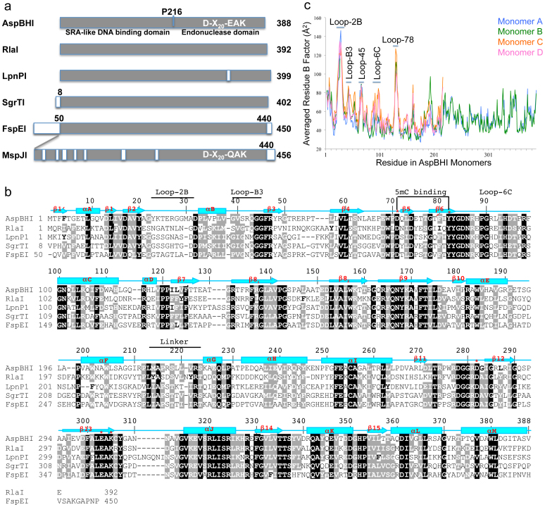
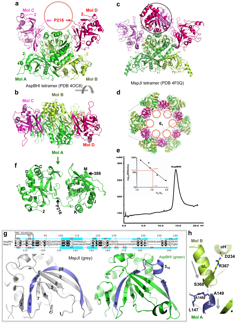
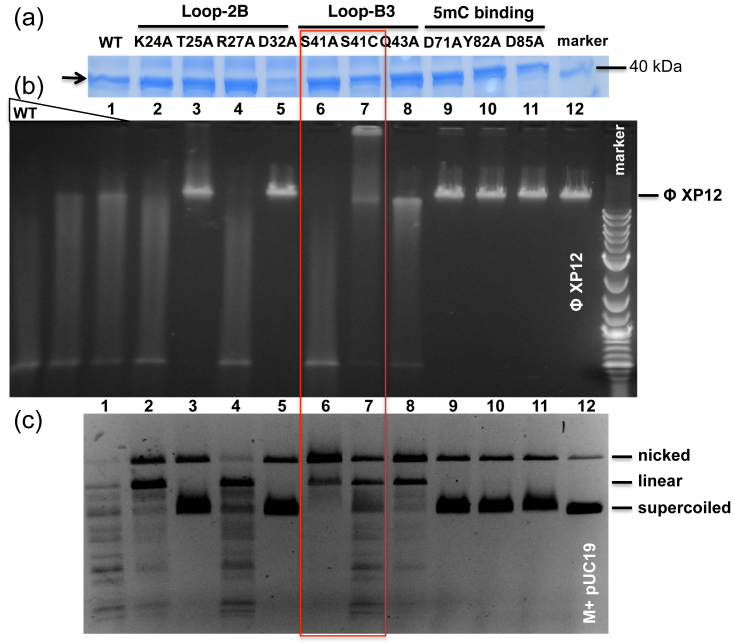
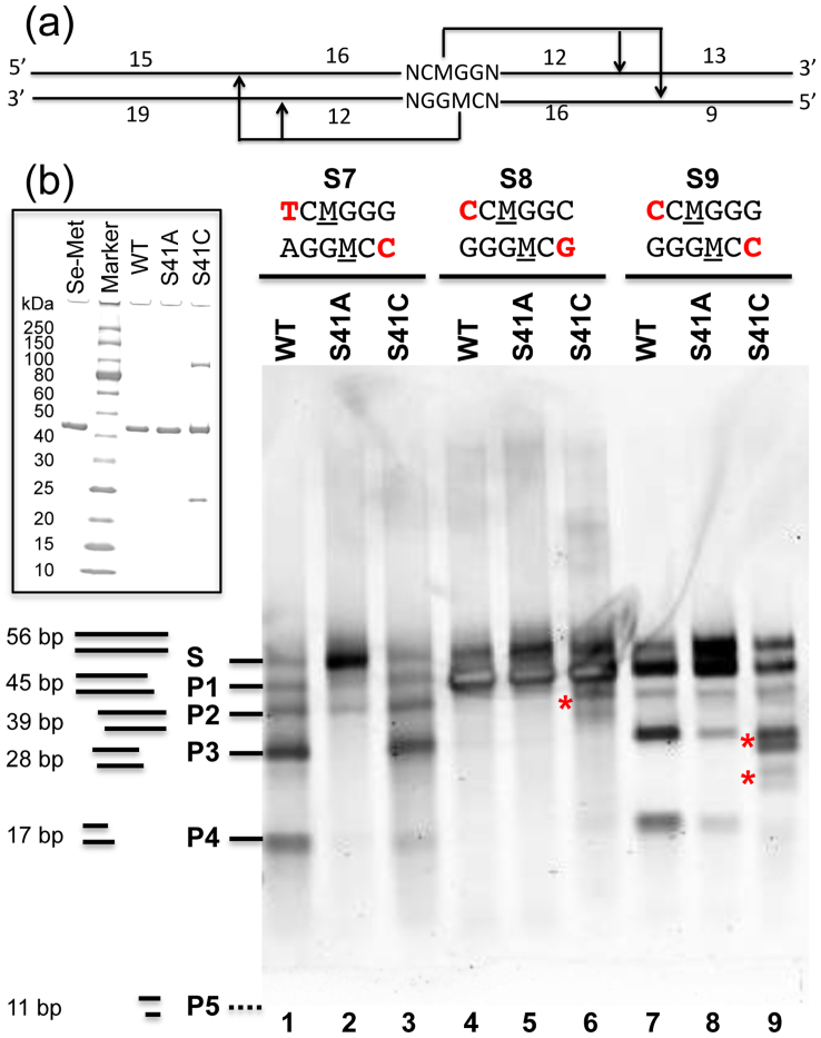
The modification-dependent restriction endonuclease AspBHI recognizes 5-methylcytosine (5mC) in the double-strand DNA sequence context of (C/T)(C/G)(5mC)N(C/G) (N = any nucleotide) and cleaves the two strands a fixed distance (N12/N16) 3' to the modified cytosine. We determined the crystal structure of the homo-tetrameric AspBHI. Each subunit of the protein comprises two domains: an N-terminal DNA-recognition domain and a C-terminal DNA cleavage domain. The N-terminal domain is structurally similar to the eukaryotic SET and RING-associated (SRA) domain, which is known to bind to a hemi-methylated CpG dinucleotide. The C-terminal domain is structurally similar to classic Type II restriction enzymes and contains the endonuclease catalytic-site motif of DX20EAK. To understand how specific amino acids affect AspBHI recognition preference, we generated a homology model of the AspBHI-DNA complex, and probed the importance of individual amino acids by mutagenesis. Ser41 and Arg42 are predicted to be located in the DNA minor groove 5' to the modified cytosine. Substitution of Ser41 with alanine (S41A) and cysteine (S41C) resulted in mutants with altered cleavage activity. All 19 Arg42 variants resulted in loss of endonuclease activity.
Conflict of interest statement
Restriction enzymes, and modification-dependent restriction enzymes, mentioned in this article are products of New England Biolabs.
Figures

Similar articles
-
Structure of 5-hydroxymethylcytosine-specific restriction enzyme, AbaSI, in complex with DNA.Nucleic Acids Res. 2014 Jul;42(12):7947-59. doi: 10.1093/nar/gku497. Epub 2014 Jun 3. Nucleic Acids Res. 2014. PMID: 24895434 Free PMC article.
-
Structure-guided sequence specificity engineering of the modification-dependent restriction endonuclease LpnPI.Nucleic Acids Res. 2015 Jul 13;43(12):6144-55. doi: 10.1093/nar/gkv548. Epub 2015 May 22. Nucleic Acids Res. 2015. PMID: 26001968 Free PMC article.
-
Structure and cleavage activity of the tetrameric MspJI DNA modification-dependent restriction endonuclease.Nucleic Acids Res. 2012 Oct;40(19):9763-73. doi: 10.1093/nar/gks719. Epub 2012 Jul 30. Nucleic Acids Res. 2012. PMID: 22848107 Free PMC article.
-
Three-dimensional structural views of damaged-DNA recognition: T4 endonuclease V, E. coli Vsr protein, and human nucleotide excision repair factor XPA.Mutat Res. 2000 Aug 30;460(3-4):257-75. doi: 10.1016/s0921-8777(00)00031-8. Mutat Res. 2000. PMID: 10946233 Review.
-
Crystallographic and bioinformatic studies on restriction endonucleases: inference of evolutionary relationships in the "midnight zone" of homology.Curr Protein Pept Sci. 2003 Oct;4(5):327-37. doi: 10.2174/1389203033487072. Curr Protein Pept Sci. 2003. PMID: 14529527 Review.
Cited by
-
Recognition and cleavage of 5-methylcytosine DNA by bacterial SRA-HNH proteins.Nucleic Acids Res. 2015 Jan;43(2):1147-59. doi: 10.1093/nar/gku1376. Epub 2015 Jan 6. Nucleic Acids Res. 2015. PMID: 25564526 Free PMC article.
-
DNA Base Flipping: A General Mechanism for Writing, Reading, and Erasing DNA Modifications.Adv Exp Med Biol. 2016;945:321-341. doi: 10.1007/978-3-319-43624-1_14. Adv Exp Med Biol. 2016. PMID: 27826845 Free PMC article. Review.
-
Protein Domain Guided Screen for Sequence Specific and Phosphorothioate-Dependent Restriction Endonucleases.Front Microbiol. 2020 Aug 18;11:1960. doi: 10.3389/fmicb.2020.01960. eCollection 2020. Front Microbiol. 2020. PMID: 33013736 Free PMC article.
-
Structure of 5-hydroxymethylcytosine-specific restriction enzyme, AbaSI, in complex with DNA.Nucleic Acids Res. 2014 Jul;42(12):7947-59. doi: 10.1093/nar/gku497. Epub 2014 Jun 3. Nucleic Acids Res. 2014. PMID: 24895434 Free PMC article.
-
Structure-guided sequence specificity engineering of the modification-dependent restriction endonuclease LpnPI.Nucleic Acids Res. 2015 Jul 13;43(12):6144-55. doi: 10.1093/nar/gkv548. Epub 2015 May 22. Nucleic Acids Res. 2015. PMID: 26001968 Free PMC article.
References
Publication types
MeSH terms
Substances
Associated data
- Actions
Grants and funding
LinkOut - more resources
Full Text Sources
Other Literature Sources
Molecular Biology Databases
