. 2014 Sep;234(1):11-22.
doi: 10.1002/path.4349. Epub 2014 Jun 18.

Endothelial cells promote stem-like phenotype of glioma cells through activating the Hedgehog pathway

Affiliations
Free PMC article

Endothelial cells promote stem-like phenotype of glioma cells through activating the Hedgehog pathway

Guang-Ning Yan et al. J Pathol. 2014 Sep.
Free PMC article

Abstract

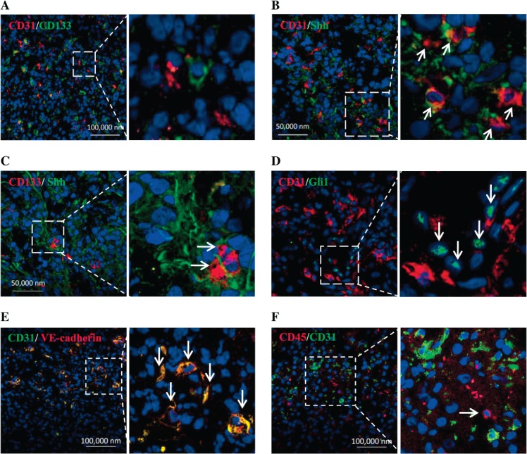
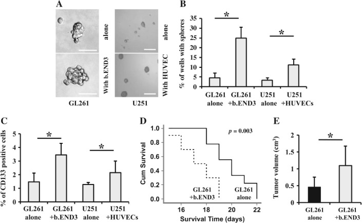
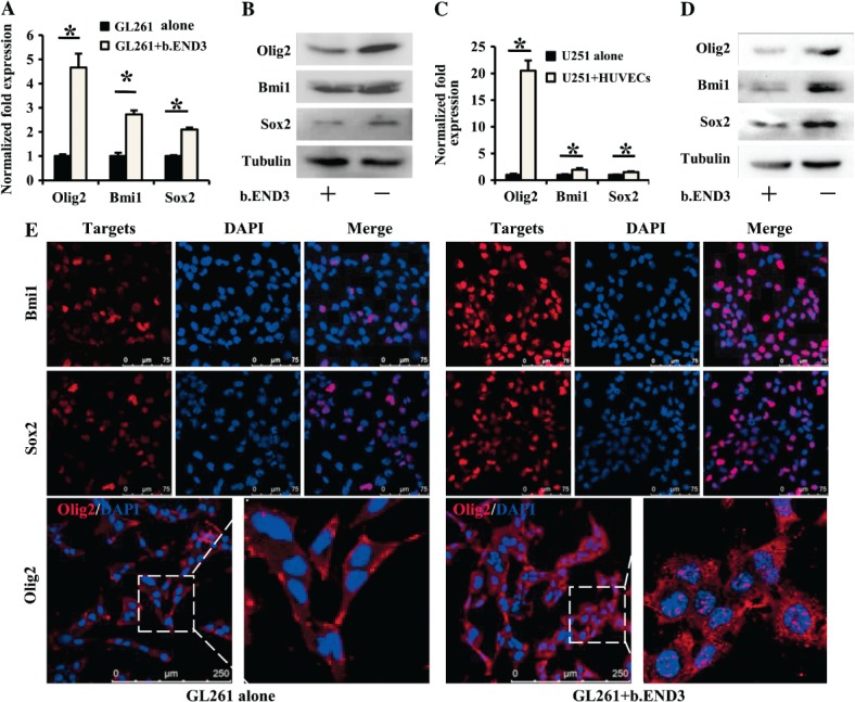
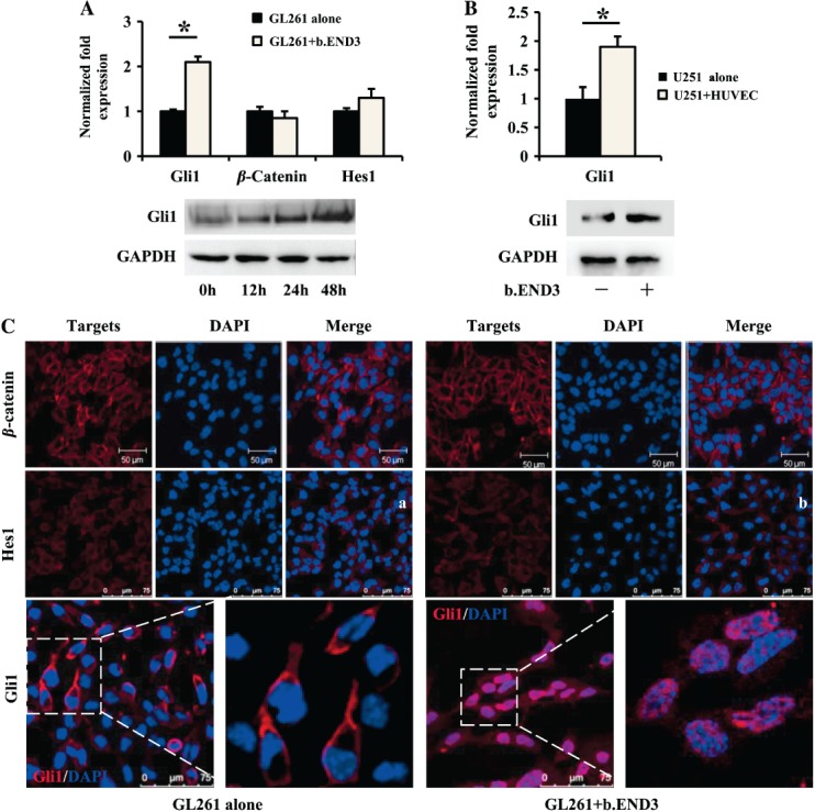
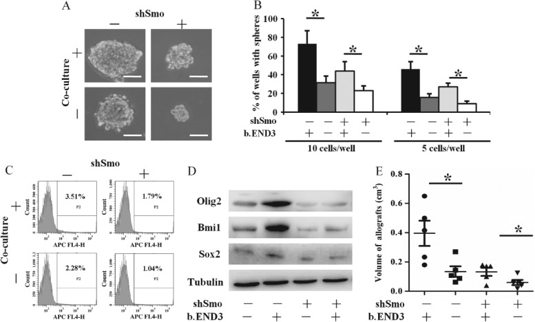
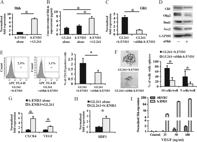
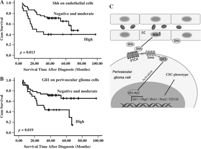
Microenvironmental regulation of cancer stem cells (CSCs) strongly influences the onset and spread of cancer. The way in which glioma cells interact with their microenvironment and acquire the phenotypes of CSCs remains elusive. We investigated how communication between vascular endothelial cells and glioma cells promoted the properties of glioma stem cells (GSCs). We observed that CD133(+) GSCs were located closely to Shh(+) endothelial cells in specimens of human glioblastoma multiforme (GBM). In both in vitro and in vivo studies, we found that endothelial cells promoted the appearance of CSC-like glioma cells, as demonstrated by increases in tumourigenicity and expression of stemness genes such as Sox2, Olig2, Bmi1 and CD133 in glioma cells that were co-cultured with endothelial cells. Knockdown of Smo in glioma cells led to a significant reduction of their CSC-like phenotype formation in vitro and in vivo. Endothelial cells with Shh knockdown failed to promote Hedgehog (HH) pathway activation and CSC-like phenotype formation in co-cultured glioma cells. By examination of glioma tissue specimens from 65 patients, we found that the survival of glioma patients was closely correlated with the expression of both Shh by endothelial cells and Gli1 by perivascular glioma cells. Taken together, our study demonstrates that endothelial cells in the tumour microenvironment provide Shh to activate the HH signalling pathway in glioma cells, thereby promoting GSC properties and glioma propagation.

Keywords: endothelial cell; glioma stem cell; hedgehog pathway; niche.


Figures

Figure 1
Figure 1
CD133-expressing GSCs locate closely to Shh-expressed endothelial cells in human GBM samples. (A) CD133+ tumour cells (green) located close to CD31+ endothelial cells (red). (B) CD31+ endothelial cells expressed Shh (orange, arrow). (C) CD133+ tumour cells (red, arrow) located next to Shh-expressed cells (green). (D) Gli1-expressed tumour cells (green, arrow) closely located to CD31+ endothelial cells (red). (E) CD31 and VE-cadherin were co-expressed in endothelial cells (yellow). (F) CD31+ endothelial cells (green) were not CD45+ (red, arrow). Right panels are partial enlargements of the corresponding left panels.
Figure 2
Figure 2
Endothelial cells promote tumour sphere formation and CD133 expression by glioma cells in vitro and tumour growth in vivo. (A) Tumour spheres formed by GL261 cells cultured with b.END3 cells (left; scale bar = 100 µm) or by U251 cells cultured with HUVECs (right; scale bar = 300 µm) were larger in size than those formed by GL261, or U251 cells alone, respectively. (B) Efficiency of tumour sphere formation by GL261 cells cultured with b.END3 cells or by U251 cells cultured with HUVECs was higher than that by GL261 or U251 cells alone, respectively (*p < 0.05). (C) The numbers of CD133+ GL261 (left) or U251 cells (right) were significantly increased after co-culture with b.END3 cells or HUVECs, respectively (*p < 0.05). (D) Survival times of mice that were orthotopically co-injected with GL261 and b.END3 cells were shorter than those of mice orthotopically injected with GL261 cells alone (*p < 0.05). (E) Tumour volume of orthotopic allografts generated by co-injection of GL261 with b.END3 cells was significantly larger than that of orthotopic allografts generated by injection of GL261 cells alone (*p < 0.05).
Figure 3
Figure 3
Endothelial cells up-regulate expression of CSC-associated genes in glioma cells. As compared to GL261 alone, expression of GSC-associated genes Olig2, Bmi1 and Sox2 were elevated in GL261 cells co-cultured with b.END3 cells: (A) real-time RT–PCR, *p < 0.05; (B) western blot, tubulin was used as control. As compared to U251 cells alone, expressions of Olig2, Bmi1 and Sox2 were elevated in U251 cells co-cultured with HUVECs; (C) real-time RT–PCR, *p < 0.05; (D) western blot, tubulin was used as control. (E) Immunofluorescence staining revealed that expressions of Bmi1 (upper), Sox2 (middle) and Olig2 (lower) were increased in GL261 cells co-cultured with b.END3 cells. Right panels are partial enlargements of the corresponding left panels.
Figure 4
Figure 4
The HH pathway is significantly activated in glioma cells co-cultured with endothelial cells. (A) Gli1 expression was induced in GL261 cells co-cultured with b.END3, while Hes1 and β-catenin were not affected (*p < 0.05; upper panel, RT–PCR; lower panel, western blot). (B) Gli1 expression was induced in U251 cells co-cultured with HUVECs (*p < 0.05; upper panel, RT–PCR; lower panel, western blot). (C) Location and fluorescence intensity of both β-catenin (upper panel) and Hes1 (middle panel) were not significantly changed. Gli1 (red) was up-regulated and translocated to the nuclei of GL261 cells co-cultured with b.END3 (lower right panel). Right panels are partial enlargements of the corresponding left panels.
Figure 5
Figure 5
Inhibition of Hedgehog pathway hampers the appearance of GSC-like phenotype of glioma cells. As compared to mock-GL261 alone or mock-GL261 cells co-cultured with b.END3, shSmo–GL261 alone or shSmo–GL261 cells co-cultured with b.END3 cells formed smaller tumour spheres (A; scale bar = 100 µm) and fewer tumour spheres (B; *p < 0.05), respectively. CD133 expression of shSmo–GL261 cells was reduced (C) and expressions of Sox2, Olig2 and Bmi1 were significantly reduced (D), respectively, tubulin was used as control. Subcutaneous allografts generated by shSmo–GL261 cells, with or without b.END3 cells, were significantly smaller in volume as compared to mock GL261 cells with or without b.END3 cells (E; *p < 0.05), respectively; −, wild-type GL261 cells; +, Smo-knockdown GL261 cells.
Figure 6
Figure 6
Shh secreted from endothelial cells is important in both activating HH pathway and promoting CSC-like phenotype in glioma cells. As compared to b.END3 cells alone, mRNA expression of Shh in b.END3 (A, *p < 0.05) and secretion of Shh protein in supernatant (B, *p < 0.05) were significantly up-regulated when b.END3 was co-cultured with GL261 cells. By comparison with GL261 cells that were co-cultured with wild-type b.END3 cells, Shh knockdown in b.END3 cells (siShh-b.END3) inhibited expression of Gli1, Olig2, Sox2 and Bmi1 in GL261 cells (C; RT–PCR; D; western blot; *p < 0.05). The CD133 expression (E; *p < 0.05) and tumour spheres (F; *p < 0.05; scale bar = 50 µm) of GL261 cells cultured with siShh-b.END3 cells were significantly less than those co-cultured with wild-type b.END3 cells. Both CXCR4 and VEGF in b.END3 cells (G; *p < 0.05) and SDF1 in GL261 cells (H; *p < 0.05) were induced after co-culture of GL261 cells and b.END3 cells, respectively. VEGF protein treatment induced Shh expression in both b.END3 cells and HUVECs (I; *p < 0.05); −, GL261 cells cultured with wild-type b.END3 cells; +, GL261 cells cultured with Shh-knockdown b.END3 cells.
Figure 7
Figure 7
Prognosis of glioma patients is associated with Shh expression by endothelial cells and Gli1 by glioma cells. (A) Kaplan–Meier curve showing higher expression of Shh by endothelial cells significantly relates to worse prognosis. (B) Kaplan–Meier curve showing that higher expression of Gli1 by perivascular glioma cells significantly relates to worse prognosis. (C) Schematic presentation for summarization.

References

    1. Li J, Di C, Mattox AK, et al. The future role of personalized medicine in the treatment of glioblastoma multiforme. Pharmgenom Pers Med. 2010;3:111–127. - PMC - PubMed
    1. Welte Y, Adjaye J, Lehrach HR, et al. Cancer stem cells in solid tumors: elusive or illusive? Cell Commun Signal. 2010;8(1):6. - PMC - PubMed
    1. Rapp UR, Ceteci F. Schreck R. Oncogene-induced plasticity and cancer stem cells. Cell Cycle. 2008;7(1):45–51. - PubMed
    1. Heddleston JM, Li Z, McLendon RE, et al. The hypoxic microenvironment maintains glioblastoma stem cells and promotes reprogramming towards a cancer stem cell phenotype. Cell Cycle. 2009;8(20):3274–3284. - PMC - PubMed
    1. Moore KA. Stem cells and their niches. Science. 2006;311:1880–1885. - PubMed

Publication types

MeSH terms