. 2014 May;33(5):1075-82.
doi: 10.3892/ijmm.2014.1687. Epub 2014 Mar 6.

Islet-1 promotes the cardiac-specific differentiation of mesenchymal stem cells through the regulation of histone acetylation

Affiliations

Islet-1 promotes the cardiac-specific differentiation of mesenchymal stem cells through the regulation of histone acetylation

Naijing Yin et al. Int J Mol Med. 2014 May.

Abstract

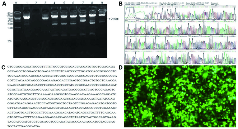
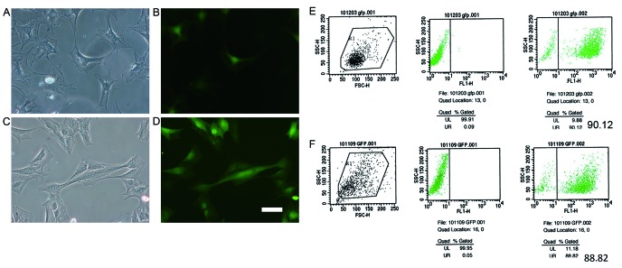
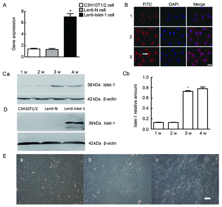
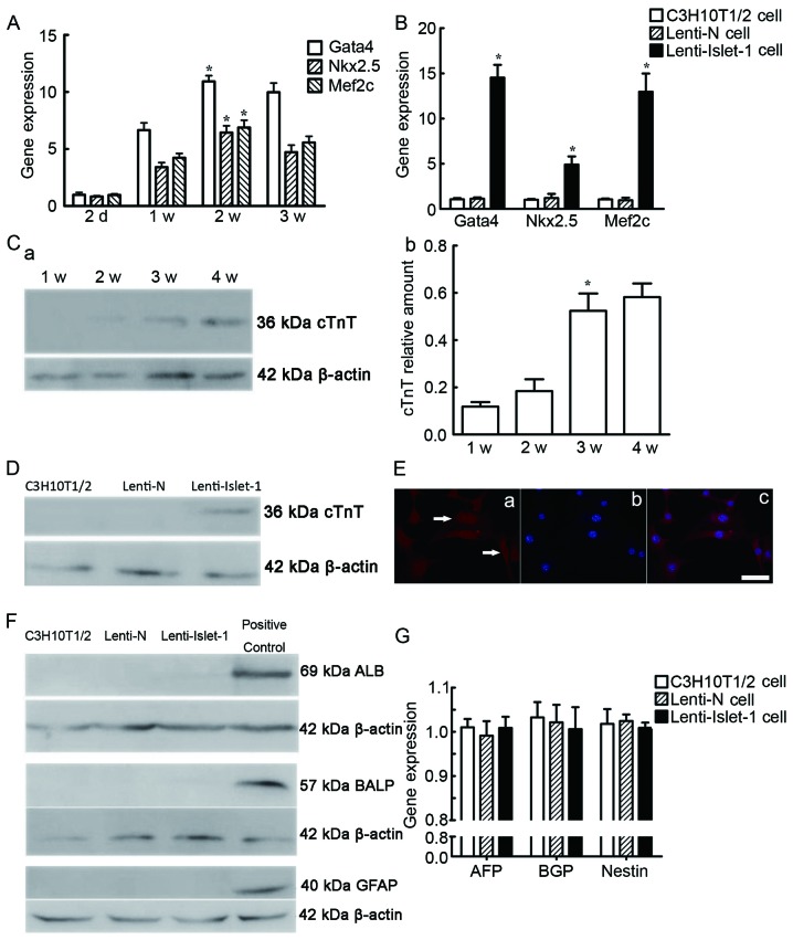
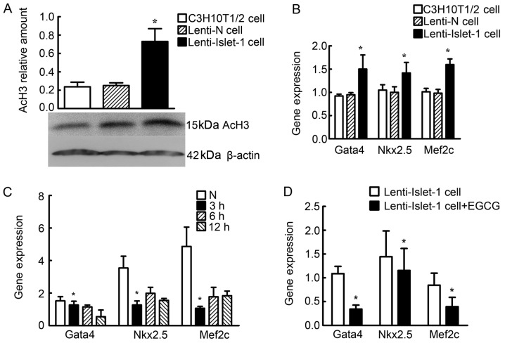
The aim of the present study was to investigate the effects of Islet-1 on the process of mesenchymal stem cell (MSC) differentiation into cardiomyocyte-like cells and to elucidate the possible mechanisms involved. Lentiviral vectors expressing Islet-1 (Lenti-Islet-1) were constructed and used for C3H10T1/2 cell transfection. Cell morphology was observed. Cardiac-related genes and proteins were detected by qPCR and western blot analysis. Epigallocatechin gallate (EGCG) was used as an inhibitor of acetylated histone H3 (AcH3). AcH3 was detected by chromatin immunoprecipitation. Cells overexpressing Islet-1 tended to change into fibroblast-like cells and were arranged in the same direction. The enhanced expression of GATA binding protein 4 (Gata4), NK2 homeobox 5 (Nkx2.5), myocyte enhancer factor 2C (Mef2c) and cardiac troponin T (cTnT) was observed in the cells overexpressing Islet-1 following transfection with Lenti-Islet-1. However, the expression of hepatocyte-, bone- and neuronal-specific markers was not affected by Islet-1. The AcH3 relative amount increased following transfection with Lenti-Islet-1, which was associated with the enhanced expression of Gata4, Nkx2.5 and Mef2c in these cells. The expression of Gata4, Nkx2.5 and Mef2c in the C3H10T1/2 cells transfected with Lenti-Islet-1 and treated with EGCG was reduced following treatment with EGCG. The data presented in this study indicate that Islet-1 specifically induces the differentiation of C3H10T1/2 cells into cardiomyocyte-like cells, and one of the mechanisms involved is the regulation of histone acetylation.


Figures

Figure 1
Figure 1
(A) Detection of lentiviral vector. PCR of random clones. Lane M, DL15000 DNA marker; lanes 1–9, clones that we selected. The 7th lane shows the positive clone. (B and C) Part of the Lenti-Islet-1 plasmid sequencing result. (D) Detection of green fluorescent protein (GFP) expression in 293T cells following transfection with Lenti-Islet-1 vectors (magnification, ×10). Scale bar, 100 μm.
Figure 2
Figure 2
Green fluorescent protein (GFP) expression detected under a fluorescence microscope. The lentiviral vectors carried the GFP gene; thus, a fluorescence microscope was used to detected GFP expression in the C3H10T1/2 cells transfected with Lenti-N and the C3H10T1/2 cells transfected with Lenti-Islet-1 at 3 days after transfection. (A) Cell morphology of the C3H10T1/2 cells transfected with Lenti-N observed under a microscope (magnification, ×20). (B) GFP expression of the C3H10T1/2 cells transfected with Lenti-N observed udner a fluorescence microscope (magnification, ×20). (C) Cell morphology of the C3H10T1/2 cells transfected with Lenti-Islet-1 observed under a microscope (magnification, ×20). (D) Cell morphology of the C3H10T1/2 cells transfected with Lenti-Islet-1 observed under a microscope (magnification, ×20). Scale bar, 20 μm. (E and F) Transfection efficiency of teh C3H10T1/2 cells transfected with Lenti-N and the C3H10T1/2 cells transfected with Lenti-Islet-1 detected by flow cytometry (FCM). The transfection efficiency of the C3H10T1/2 cells transduced with lentiviral vectors with pWPI-GFP plasmid or lentiviral vectors with pWPI-GFP-Islet-1 plasmid was detected by FCM. (E) The transfection efficiency of the C3H10T1/2 cells transfected with Lenti-N was 90.12%. (F) The transfection efficiency of the C3H10T1/2 cells transfected with Lenti-Islet-1 was 88.82%.
Figure 3
Figure 3
(A) Islet-1 gene expression in the C3H10T1/2 cells transfected with Lenti-Islet-1 was higher (*P<0.05) than that in the other C3H10T1/2 cells. (B) The expression of Islet-1 was detected by immunofluorescence in each group: Panel 1, untransfected C3H10T1/2 cells; panel 2, C3H10T1/2 cells transfected with Lenti-N; panel 3, C3H10T1/2 cells transfected with Lenti-Islet-1. FITC staining of Islet-1 protein showing its location. DAPI staining of cell nucleus. Merge image was obtained by overlapping FITC and DAPI images (magnification, ×20). Scale bar, 20 μm. (C-a) The expression of Islet-1 at different time points in the C3H10T1/2 cells transfected with Lenti-Islet-1: 1w, 1st week; 2w, 2nd week; 3w, 3rd week; 4w, 4th week. (C-b) Islet-1 protein expression in the C3H10T1/2 cells transfected with Lenti-Islet-1 was higher during the 3rd week than in the first 2 weeks (*P<0.05). (D) Islet-1 protein expression in the C3H10T1/2 cells transfected with Lenti-Islet-1 was higher than that in the untransfected C3H10T1/2 cells and the C3H10T1/2 cells transfected with Lenti-N. (E) Morphological changes of C3H10T1/2 cells transfected with lentiviral vectors with pWPI-GFP plasmid or lentiviral vectors with pWPI-GFP-Islet-1 plasmid observed under a microscope. No difference was observed between the untransfected C3H10T1/2 cells (magnification, ×10) (E-a) and the C3H10T1/2 cells transfected with Lenti-N (magnification, ×10) (E-b), whereas the C3H10T1/2 cells transfected with Lenti-Islet-1 (×10) (E-c) turned into fibroblast-like cells and were arranged toward the same direction. Scale bar, 100 μm.
Figure 4
Figure 4
(A) The expression of Gata4, Nkx2.5 and Mef2c gene at different time points in C3H10T1/2 cells transfected with Lenti-Islet-1. 2d, 2nd day; 1w, 1st week; 2w, 2nd week; 3w, 3rd week. The peak expression of cardiac-specific genes in the C3H10T1/2 cells transfected with Lenti-Islet-1 was significantly higher than that in the untransfected C3H10T1/2 cells and the C3H10T1/2 cells transfected with Lenti-N at 2 weeks after transfection (*P<0.05). (B) The expression of cardiac-specific genes in the C3H10T1/2 cells transfected with Lenti-Islet-1 was higher in the 2nd week than the other time points (*P<0.05). (C) The protein expression of cTnT at different time points in the C3H10T1/2 cells transfected with Lenti-Islet-1. 1w, 1st week; 2w, 2nd week; 3w, 3rd week; 4w, 4th week. cTnT expression increased from the 3rd week following transfection (*P<0.05). (D) cTnT expression in the untransfected C3H10T1/2 cells, the C3H10T1/2 cells transfected with Lenti-N and the C3H10T1/2 cells transfected with Lenti-Islet-1 at 3 weeks after transfection. cTnT expression was higher in the C3H10T1/2 cells transfected with Lenti-Islet-1 than the other 2 groups of cells. (E) cTnT expression in the C3H10T1/2 cells transfected with Lenti-Islet-1 was located in the cell nucleus and cytoplasm (magnification, ×20). Scale bar, 20 μm. (F) Expression of hepatocyte-, bone- and neuronal-specific markers. Albumin (ALB), bone-specific alkaline phosphatase (BALP) and glial fibrillary acidic protein (GFAP) expression was detected by western blot analysis in the untransfected C3H10T1/2 cells, the C3H10T1/2 cells transfected with Lenti-N and the C3H10T1/2 cells transfected with Lenti-Islet-1 and the positive control group. There was no ALB, BALP and GFAP expression observed in the untransfected C3H10T1/2 cells, the C3H10T1/2 cells transfected with Lenti-N and the C3H10T1/2 cells transfected with Lenti-Islet-1. (G) AFP, bone Gla protein (BGP) and nestin expression detected by qPCR. The expression of BGP, AFP and nestin was did not differ significantly between the 3 groups.
Figure 5
Figure 5
Differences in histone acetylation levels in the untransfected C3H10T1/2 cells, the C3H10T1/2 cells transfected with Lenti-N and the C3H10T1/2 cells transfected with Lenti-Islet-1. (A) Acetylated histone H3 (AcH3) was detected by western blot analysis in the untranstected C3H10T1/2 cells, the C3H10T1/2 cells transfected with Lenti-N and the C3H10T1/2 cells transfected with Lenti-Islet-1. The AcH3 relative amount in the C3H10T1/2 cells transfected with Lenti-Islet-1 was higher than that in the untransfected C3H10T1/2 cells and the C3H10T1/2 cells transfected with Lenti-N (*P<0.05). (B) Chromatin immunoprecipitation (ChIP) and qPCR were performed to reveal the acetylation levels of histone H3 on the cardiac-specific genes, GATA binding protein 4 (Gata4), NK2 homeobox 5 (Nkx2.5) and myocyte enhancer factor 2C (Mef2c), at their peak expression times (2 weeks after transfection). The expression of Gata4, Nkx2.5 and Mef2c combined with AcH3 in the C3H10T1/2 cells transfected with Lenti-Islet-1 was higher than that in the untransfected C3H10T1/2 cells and the C3H10T1/2 cells transfected with Lenti-N (*P<0.05). (C) Gata4, Nkx2.5 and Mef2c expression was found to be reduced 3 h following treatment with 120 μmol/l epigallocatechin gallate (EGCG) (*P<0.05). (D) Gata4, Nkx2.5 and Mef2c expression in the C3H10T1/2 cells transfected with Lenti-Islet-1 and treated with EGCG was lower compared with the cells not treated with EGCG (*P<0.05).

Similar articles

Cited by

References

    1. Christoforou N, Gearhart JD. Stem cells and their potential in cell-based cardiac therapies. Prog Cardiovasc Dis. 2007;49:396–413. - PubMed
    1. Bianco P, Robey PG, Simmons PJ. Mesenchymal stem cells: revisiting history, concepts, and assays. Cell Stem Cell. 2008;2:313–319. - PMC - PubMed
    1. Pittenger MF, Mackay AM, Beck SC, et al. Multilineage potential of adult human mesenchymal stem cells. Science. 1999;284:143–147. - PubMed
    1. Qin JJ, Xian SX, Huang XW, Sun JH. Differentiation of mesenchymal stem cells into cardiomyocyte-like cells in vitro: Drug, microenvironment and method. Zhongguo Zuzhi Gongcheng Yanjiu Yu Linchuang Kangfu. 2011;15:139–142. (In Chinese)
    1. Mafi1 P, Hindocha S, Mafi R, Griffin M, Khan WS. Adult mesenchymal stem cells and cell surface characterization - a systematic review of the literature. Open Orthop J. 2011;5:253–260. - PMC - PubMed

Publication types

MeSH terms