Review
. 2014 Nov;229(11):1584-94.
doi: 10.1002/jcp.24604.

Ribosome biogenesis: emerging evidence for a central role in the regulation of skeletal muscle mass

Affiliations
Review

Ribosome biogenesis: emerging evidence for a central role in the regulation of skeletal muscle mass

Thomas Chaillou et al. J Cell Physiol. 2014 Nov.

Abstract

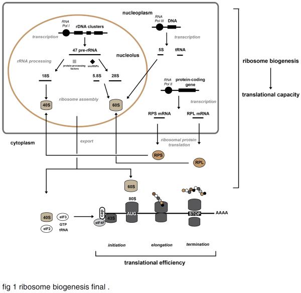
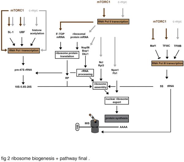
The ribosome is a supramolecular ribonucleoprotein complex that functions at the heart of the translation machinery to convert mRNA into protein. Ribosome biogenesis is the primary determinant of translational capacity of the cell and accordingly has an essential role in the control of cell growth in eukaryotes. Cumulative evidence supports the hypothesis that ribosome biogenesis has an important role in the regulation of skeletal muscle mass. The purpose of this review is to, first, summarize the main mechanisms known to regulate ribosome biogenesis and, second, put forth the hypothesis that ribosome biogenesis is a central mechanism used by skeletal muscle to regulate protein synthesis and control skeletal muscle mass in response to anabolic and catabolic stimuli. The mTORC1 and Wnt/β-catenin/c-myc signaling pathways are discussed as the major pathways that work in concert with each of the three RNA polymerases (RNA Pol I, II, and III) in regulating ribosome biogenesis. Consistent with our hypothesis, activation of these two pathways has been shown to be associated with ribosome biogenesis during skeletal muscle hypertrophy. Although further study is required, the finding that ribosome biogenesis is altered under catabolic states, in particular during disuse atrophy, suggests that its activation represents a novel therapeutic target to reduce or prevent muscle atrophy. Lastly, the emerging field of ribosome specialization is discussed and its potential role in the regulation of gene expression during periods of skeletal muscle plasticity.


Figures

Fig. 1
Fig. 1
Schematic representation of the cellular translation machinery. Ribosome biogenesis is a well-orchestrated process that involves the coordinated actions of the three types of RNA polymerase (RNA Pol I, -II and -III). RNA Pol I is responsible for the transcription of the polycistronic 47S pre-rRNA in the nucleolus, which is subsequently spliced and modified by small nuclear ribonucleoproteins (snoRNPs) and several protein-processing factors to form 18S, 5.8S and 28S rRNAs. The nucleoplasmic transcription of 5S rRNA and the tRNAs is achieved by RNA Pol III while the ribosomal protein encoding-genes are transcribed in the nucleoplasm by RNA Pol II. Following the translation in the cytoplasm, the proteins of the small (RPS, ribosomal protein small) and large (RPL, ribosomal protein large) ribosomal subunits are imported into the nucleolus to be assembled with their respective ribosomal subunit. The 5.8S, 28S and 5S rRNAs assemble with RPL proteins to form the 60S ribosome subunit while the 18S rRNA and the RPS proteins form the 40S ribosome subunit. The 40S and 60S subunits are then exported to the cytoplasm, where they form the mature 80S ribosome complex. The process of ribosome biogenesis is primary determinant of the translational capacity of the cell. After nuclear export, the 40S subunit interacts with the eukaryotic initiation factor (eIF) eIF3 and the ternary complex (eIF2, tRNA and GTP) to form the pre-initiation 43S complex. This complex, which is then recruited to the m7GpppN cap structure of the mRNA after its binding to eIF4F complex, scans the 5' UTR (untranslated region) of the mRNA until the initiation codon (AUG) is detected (initiation). The recruitment of 60S subunit at the initiation codon allows the formation of the 80S complex and the start of the elongation of the mRNA. Finally, the finished polypeptide is released from the ribosome when a stop codon is encountered (termination). These three steps of translation (initiation, elongation and termination) determine the translational efficiency. This schematic representation was adapted from (van Riggelen et al., 2010) and (Boisvert et al., 2007).
Fig. 2
Fig. 2
Regulation of ribosome biogenesis by mTORC1 and c-myc. mTORC1 (mechanical target of rapamycin complex 1) and c-myc (c-myelocytomatosis oncogene) work in concert with each of the three RNA polymerases in regulating ribosome biogenesis. mTORC1 and c-myc seem to directly promote the transcription of the pre-rRNA 47S, and can lead to the activation of the RNA Pol I transcriptional factor SL-1 (selectively factor-1), and the rDNA transcription factor UBF (upstream binding factor). Some lines of evidence also suggest that c-myc indirectly increases the RNA Pol I transcription by promoting the opening of the chromatin structure near rDNA loci through histone acetylation. mTORC1 appears to regulate the translation of 5'-TOP (terminal tract of pyrimidine) mRNAs which encode ribosomal proteins and other components of the translational machinery, while c-myc activates the transcription of several ribosomal protein-encoding genes. In addition, c-myc promotes the transcription of several auxiliary factors required for ribosome biogenesis, such as genes involved in rRNA processing [Nop56 (Nop56 ribonucleoprotein), Bop1 (block of proliferation 1) and Dkc1 (dyskeratosis congenital 1, dyskerin)], ribosome assembly [Ncl (nucleolin) and Rpl3 (ribosome protein L3)], and nuclear ribosome export [Npm1 (nucleophosmin 1) and Fbl (fibrillarin)]. Finally, mTORC1 and c-myc activate the RNA Pol III transcription through their interaction with TFIIIC (transcription factor IIIC) and TFIIIB (transcription factor IIIB), respectively. mTORC1 may also inhibit the function of the RNA Pol III repressor Maf1.

References

    1. Adams GR, Caiozzo VJ, Haddad F, Baldwin KM. Cellular and molecular responses to increased skeletal muscle loading after irradiation. Am J Physiol Cell Physiol. 2002;283(4):C1182–1195. - PubMed
    1. Alawi F, Lee MN. DKC1 is a direct and conserved transcriptional target of c-MYC. Biochem Biophys Res Commun. 2007;362(4):893–898. - PubMed
    1. Arabi A, Wu S, Ridderstrale K, Bierhoff H, Shiue C, Fatyol K, Fahlen S, Hydbring P, Soderberg O, Grummt I, Larsson LG, Wright AP. c-Myc associates with ribosomal DNA and activates RNA polymerase I transcription. Nat Cell Biol. 2005;7(3):303–310. - PubMed
    1. Armstrong DD, Esser KA. Wnt/beta-catenin signaling activates growth-control genes during overload-induced skeletal muscle hypertrophy. Am J Physiol Cell Physiol. 2005;289(4):C853–859. - PubMed
    1. Armstrong DD, Wong VL, Esser KA. Expression of beta-catenin is necessary for physiological growth of adult skeletal muscle. Am J Physiol Cell Physiol. 2006;291(1):C185–188. - PubMed

LinkOut - more resources