

doi: 10.4103/0971-3026.125577.
Radiological review of pleural tumors
Affiliations
- PMID: 24604935
- PMCID: PMC3932573
- DOI: 10.4103/0971-3026.125577
Item in Clipboard
Radiological review of pleural tumors
Indian J Radiol Imaging.
2013 Oct.
Abstract
Tumors of the pleura are not uncommon and diagnosis is clinched by combined imaging and clinical correlation. Malignant tumors are more common than benign tumors. Initial imaging modalities are chest radiography and Computed Tomography (CT). Further characterization may be required using Ultrasoundgraphy (USG), Magnetic resonance Imaging (MRI) and PET-CT. Biopsy remains gold standard. This article highlights various common and uncommon tumors of pleura and characteristic imaging findings.
Keywords: Fibroma; mesothelioma; nodular pleural thickening; pleura.
Conflict of interest statement
Figures
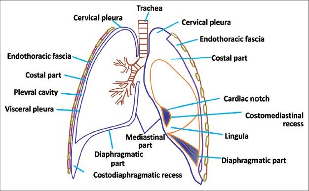
Line diagram of pleura showing different components of pleura
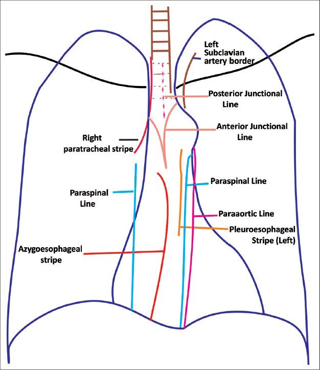
Line diagram showing junctional lines formed by pleural invaginations
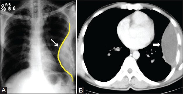
Loculated empyema: (A) Chest radiograph showing pleural-based opacity (arrow) with tapering obtuse margins in left hemithorax; (B) axial contrast-enhanced CT scan showing loculated collection (arrowhead) with peripherally enhancing thick walls
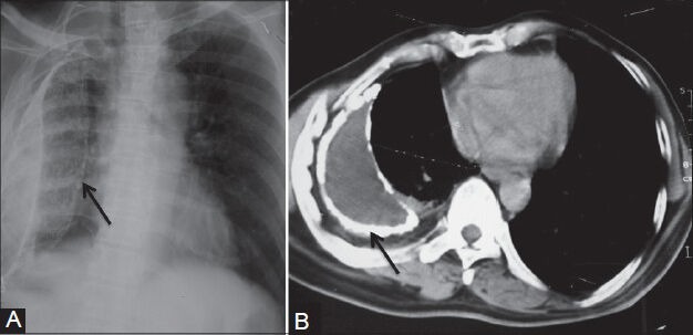
Calcified empyema: (A) Chest radiograph showing volume loss right hemithorax with veil-like calcified (arrow) pleural opacity; (B) axial contrast-enhanced CT scan showing evidence of calcified chronic empyema (arrow) with proliferation of extrapleural fat and crowding of ribs suggestive of volume loss in right hemithorax
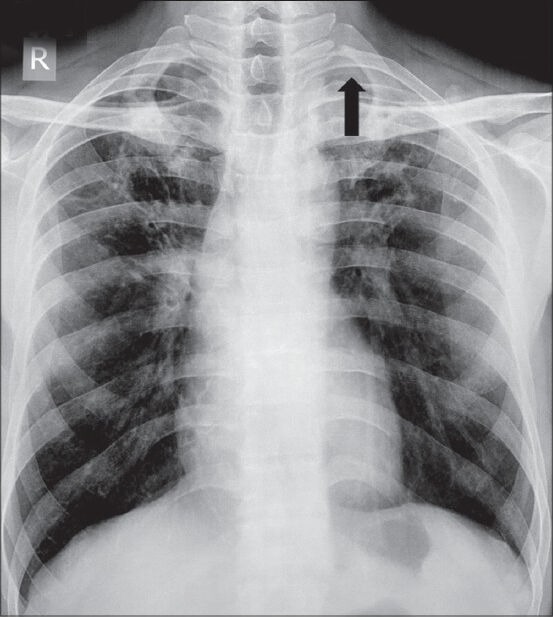
Apical pleural thickening: Chest radiograph showing apical pleural thickening (arrowhead) in left apical region
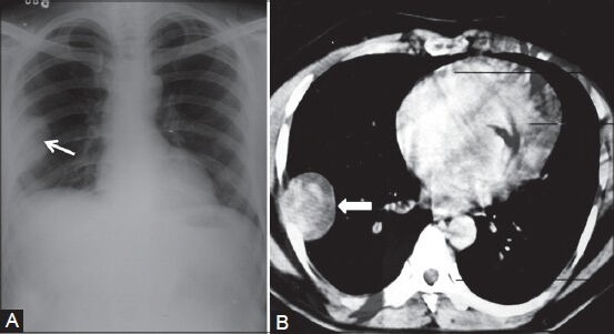
Benign solitary fibrous tumor: (A) Chest radiograph showing pleural-based opacity (arrow) in right hemithorax with peripheral obtuse margins; (B) axial contrast-enhanced CT scan showing heterogeneously enhancing pleural-based mass (arrowhead) proved to be benign fibrous pleural tumor
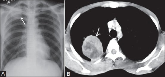
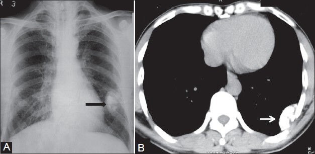
Pleural fibroma: (A) Chest radiograph showing lobulated pleural-based opacity (arrow) in right apical region; (B) axial contrast-enhanced CT scan showing heterogeneously enhancing peripheral mass lesion (arrow) in a biopsy-proven case of benign pleural fibroma
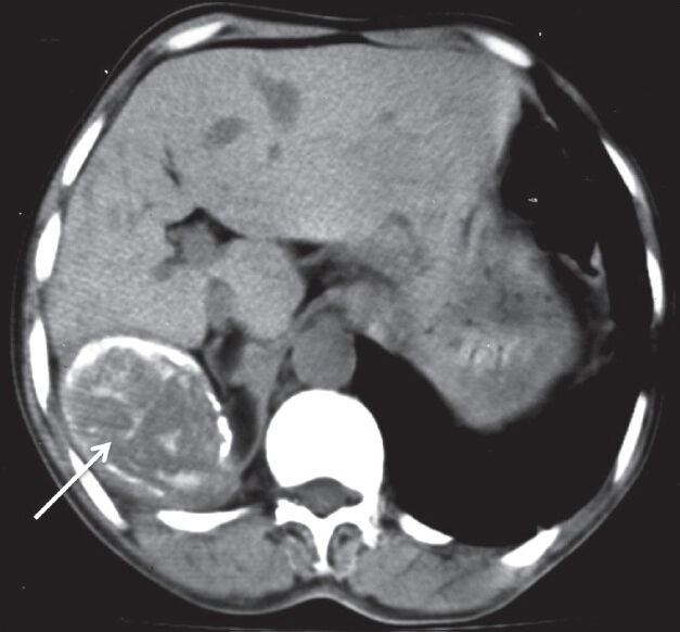
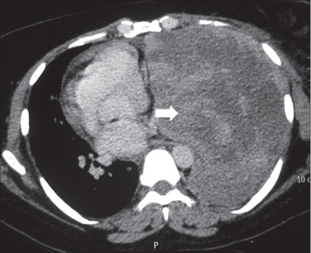
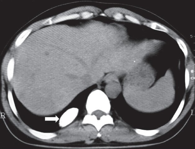
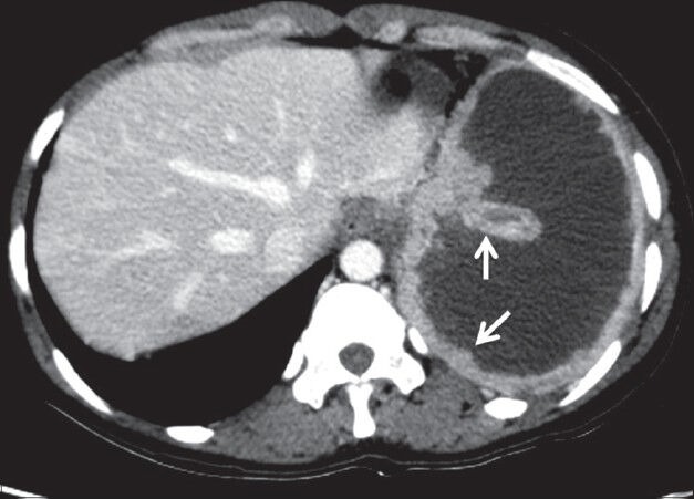
Malignant solitary fibrous tumor of pleura: Plain axial CT scan showing pleural-based soft tissue lesion with peripheral as well as internal calcification (arrow) abutting the liver
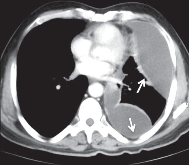
Malignant fibrous tumor of pleura: Axial contrast-enhanced CT scan showing heterogeneously enhancing mass lesion left hemithorax (arrowhead) causing mediastinal displacement to the right
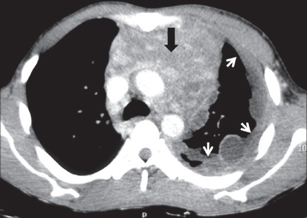
Malignant mesothelioma: Axial contrast-enhanced CT scan showing enhancing nodular pleural thickening (arrows) involving the costal and mediastinal pleura, extending into the major fissure (arrowhead) with crowding of ribs suggestive of volume loss changes in left hemithorax
Malignant mesothelioma: Axial contrast-enhanced CT scan showing homogeneously enhancing nodular pleural thickening (arrows) involving the mediastinal and costal pleura with volume loss changes in left hemithorax
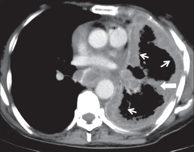
Mesothelioma presenting as pleural collections: Axial contrast-enhanced CT scan showing nodular thickening of pleura involving right hemithorax with small pleural collections (arrows)
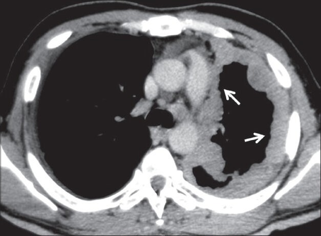
Mesothelioma presenting as a pleural effusion: Axial contrastenhanced CT scan showing moderate left pleural effusion as loculated collection with thickening of pleura (arrows) in a case of mesothelioma
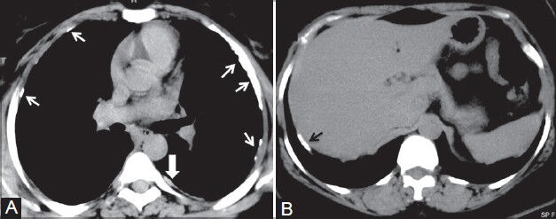
Mesothelioma and pleural plaques: (A) Axial plain CT scan showing calcified (arrows) and noncalcified (arrowhead) pleural plaques; (B) axial plain CT scan image showing calcified plaque (black arrow) classically involving the diaphragmatic parietal pleura in a construction worker
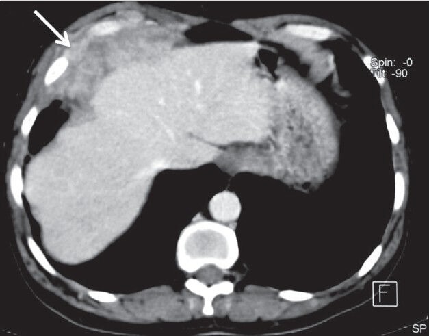
Pleural lymphoma: Axial contrast-enhanced CT scan showing heterogeneously enhancing lobulated mass lesion involving the diaphragmatic pleura (arrow) and invading the chest wall in a case of high-grade lymphoma
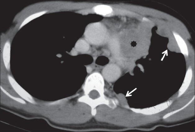
Pleural lymphoma: Axial contrast-enhanced CT scan showing homogeneously enhancing nodular pleural thickening (arrows) involving the costal pleura with mediastinal lymphadenopathy (asterisk)
Calcifying fibrous pseudotumor: (A) Chest radiograph showing pleural-based calcified opacity (arrowhead) left hemithorax with incomplete border sign; (B) plain axial CT scan image showing pleural-based calcified lesion (arrow) with no destruction of underlying ribs
Calcifying fibrous pseudotumor: Plain axial CT scan showing calcified pleural-based opacity in right hemithorax (arrowhead)
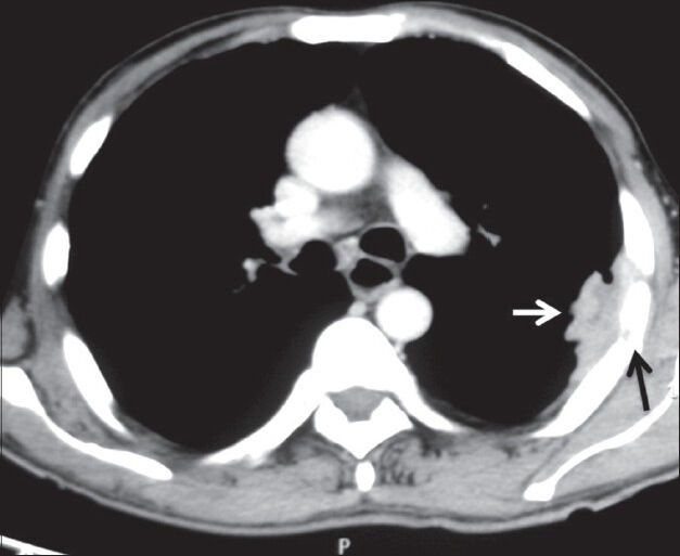
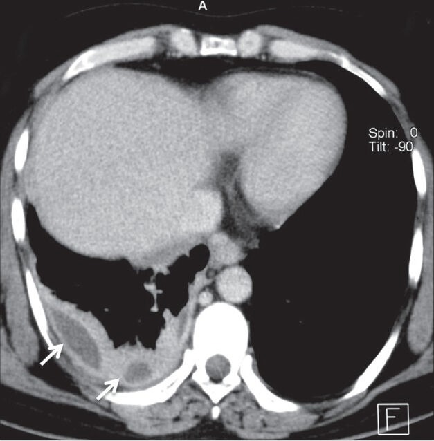
Pleural metastases: Axial contrast-enhanced CT scan showing heterogeneously enhancing pleural-based soft tissue (white arrow) with rib destruction (black arrow) in a case of pleural metastases from renal cell carcinoma
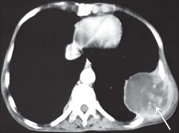
Pleural metastases: Axial contrast-enhanced CT scan showing heterogeneously enhancing pleural-based mass lesion (arrow) in left hemithorax with extrathoracic extension in a case of metastatic adenocarcinoma
Pleural metastases: Axial contrast-enhanced CT scan showing nodular pleural thickening (arrows) involving the costal and mediastinal pleura with malignant pleural effusion in a case of metastatic ovarian adenocarcinoma
Pleural drop metastases in invasive thymoma: Axial contrast-enhanced CT image showing heterogeneously enhancing anterior mediastinal mass (arrowhead) with mild left pleural effusion and ipsilateral pleural implants (arrows)
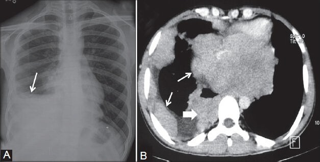
Askin tumor: (A) Chest radiograph showing inhomogeneous opacity (arrow) right hemithorax obscuring right hemidiaphragm without mediastinal shift; (B) axial contrast-enhanced CT scan showing heterogeneously enhancing nodular pleural-based lesions (arrows) involving the costal and mediastinal pleura with characteristic involvement of the sympathetic chain (arrowhead) in right paraspinal region
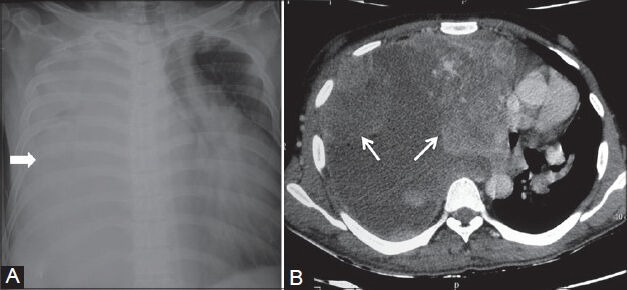
Spindle cell sarcoma of pleura: (A) Chest radiograph showing complete opacification of right hemithorax (arrowhead) with mediastinal shift to the left; (B) axial contrast-enhanced CT scan showing heterogeneously enhancing nodular pleural-based lesions with pleural effusion displacing the heart to the left
References
-
- Downer NJ, Ali NJ, Au-Yong IT. Investigating pleural thickening. BMJ. 2013;346:e8376. - PubMed
-
- Jelen TH, Bankier AA, Eisenberg RL. Solid Pleural Lesions. Am J Roentgenol. 2012;198:W512–20. - PubMed
-
- Alavi A, Gupta N, Alberini JL, Hickeson M, Adam LE, Bhargava P, et al. Positron emission tomography imaging in nonmalignant thoracic disorders. Semin Nucl Med. 2002;32:293–321. - PubMed
-
- Makis W, Ciarallo A, Hickeson M, Rush C, Novales-Diaz JA, Derbekyan V, et al. Spectrum of malignant pleural and pericardial disease on FDG PET/CT. AJR Am J Roentgenol. 2012;198:678–85. - PubMed
-
- Cardillo G, Facciolo F, Cavazzana AO, Capece G, Gasparri R, Martelli M. Localized (solitary) fibrous tumors of the pleura: An analysis of 55 patients. Ann Thorac Surg. 2000;70:1808–12. - PubMed
LinkOut - more resources
Full Text Sources
Other Literature Sources
Medical