

Cyclin D1 expression in multiple myeloma by immunohistochemistry: Case series of 14 patients and literature review
- PMID: 24604959
- PMCID: PMC3932597
- DOI: 10.4103/0971-5851.125246
Cyclin D1 expression in multiple myeloma by immunohistochemistry: Case series of 14 patients and literature review
Abstract
Background: Cyclin D1 dysregulation is an early and unifying oncogenic event in patients of multiple myeloma (MM). This may be detected up to 30% cases by immunohistochemistry (IHC) and up to 40-50% cases by molecular studies. However, studies on the clinical significance of cyclin D1 dysregulation in MM have been inconclusive. We aimed to study the pattern of cyclin D1 expression in MM by IHC and correlate with selected clinicopathologic features.
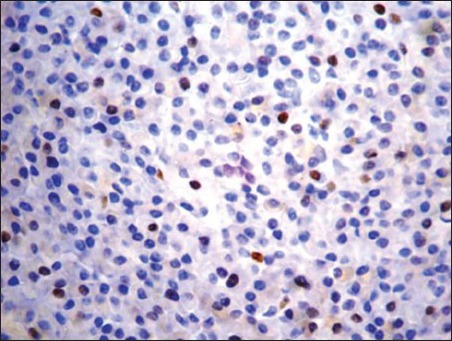
Materials and methods: Formalin fixed, decalcified, bone marrow trephine sections from 14 symptomatic patients of MM (13 newly diagnosed and one relapsed) were subjected to cyclin D1 IHC by using a rabbit monoclonal antibody to cyclin D1 (clone EPR2241).
Results: Cyclin D1 expression (in ≥10% tumor cell nuclei) was observed in 8 of 14 cases (57%). Cyclin D1 positive (+) group had significantly lower hemoglobin level (P = 0.03) than cyclin D1 negative (-) group (n = 6); though both groups showed no statistical significance (P > 0.05) in regard to age, gender, Durie and Salmon stage, lytic bone lesions, light chain phenotype, creatinine, calcium, lactate dehydrogenase, leukocyte and platelet count and bone marrow histology. Ten of 14 (71.5%) showed a favorable response (follow-up; 7 days to 34 months) to thalidomide and/or bortezomib based chemotherapeutic regimen. Four of eight cyclin D1- patients showed complete response, two had a partial response (PR) and two died of the disease; whereas 4/6 cyclin D1 - patients had PR, one refused definitive therapy and one was lost to follow-up (P > 0.05, Fischer's exact test).
Conclusion: IHC may be a feasible tool for the demonstration of cyclin D1 expression on adequately processed trephine biopsy specimen in MM patients in a resource poor setting. Negative IHC results should be correlated with molecular techniques for prognostication.
Keywords: Bone marrow; cyclin D1; immunohistochemistry; multiple myeloma; prognosis.
Conflict of interest statement
Figures


References
-
- Palumbo A, Anderson K. Multiple myeloma. N Engl J Med. 2011;364:1046–60. - PubMed
-
- McKenna RW, Kyle RA, Kuehl WM, Grogan TM, Harris NL, Coupland RW. Plasma cell neoplasm. In: Swerdlow SH, Campo E, Harris NL, Jaffe ES, Pileri SA, Stein H, et al., editors. WHO Classification of Tumors of Hematopoietic and Lymphoid Tissues. 4th ed. Lyon: IARC; 2008. pp. 200–12.
-
- International Myeloma Working Group. Criteria for the classification of monoclonal gammopathies, multiple myeloma and related disorders: A report of the international myeloma working group. Br J Haematol. 2003;121:749–57. - PubMed
LinkOut - more resources
Full Text Sources
Other Literature Sources
Research Materials
