Review
. 2014 Mar;6(1):1-8.
doi: 10.4055/cios.2014.6.1.1. Epub 2014 Feb 14.

Gap balancing vs. measured resection technique in total knee arthroplasty

Affiliations
Review

Gap balancing vs. measured resection technique in total knee arthroplasty

Brian K Daines et al. Clin Orthop Surg. 2014 Mar.

Abstract

A goal of total knee arthroplasty is to obtain symmetric and balanced flexion and extension gaps. Controversy exists regarding the best surgical technique to utilize to obtain gap balance. Some favor the use of a measured resection technique in which bone landmarks, such as the transepicondylar, the anterior-posterior, or the posterior condylar axes are used to determine proper femoral component rotation and subsequent gap balance. Others favor a gap balancing technique in which the femoral component is positioned parallel to the resected proximal tibia with each collateral ligament equally tensioned to obtain a rectangular flexion gap. Two scientific studies have been performed comparing the two surgical techniques. The first utilized computer navigation and demonstrated a balanced and rectangular flexion gap was obtained much more frequently with use of a gap balanced technique. The second utilized in vivo video fluoroscopy and demonstrated a much high incidence of femoral condylar lift-off (instability) when a measured resection technique was used. In summary, the authors believe gap balancing techniques provide superior gap balance and function following total knee arthroplasty.

Keywords: Gap balancing; Measured resection; Total knee arthroplasty technique.


Conflict of interest statement

No potential conflict of interest relevant to this article was reported.

Figures

Fig. 1
Fig. 1
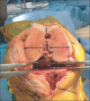
Intraoperative photograph of a flexion gap tensioning jig placed into the flexion gap, tensioning the gap, and positioned parallel to the transepicondylar axis before the anterior and posterior femoral resections are performed (Courtesy of Robert E. Booth, MD).
Fig. 2
Fig. 2
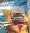
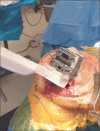
Intraoperative photograph demonstrating a tensioning jig placed into the extension gap with an extramedullary guide used to determine alignment and width of the distal femoral resection (Courtesy of Robert E. Booth, MD).
Fig. 3
Fig. 3
(A) Intraoperative photograph demonstrating resection of 4 mm of the posterior aspect of the medial femoral condyle to gain access to posterior compartment osteophytes. (B) Photograph demonstrating how an osteotome with greater curvature allows for easier access to posterior femoral osteophytes than a traditional curved osteotome. (C) Intraoperative photograph demonstrating removal of posterior femoral osteophytes using a curved osteotome.
Fig. 4
Fig. 4
Intraoperative photograph demonstrating assessment of lower extremity alignment and extension gap balance using a spacer block.
Fig. 5
Fig. 5
Intraoperative photograph of the knee at 90 degrees of flexion and the collateral ligaments equally tensioned using laminar spreaders. Note the transepicondylar axis is parallel and anterior-posterior axis is perpendicular to the resected proximal tibia.
Fig. 6
Fig. 6
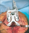
Intraoperative photograph demonstrating placement of the anteroposterior femoral cutting block parallel to the resected proximal tibia with each collateral ligament tensioned to create a rectangular flexion gap.
Fig. 7
Fig. 7
Intraoperative photograph demonstrating placement of a spacer block (same width as utilized in creation of the extension gap) into the flexion gap to assure appropriate width before performing the anterior and posterior femoral condylar resections.
Fig. 8
Fig. 8
Diagram demonstrating the transepicondylar (TEA), anterior-posterior (AP), and posterior condylar (PCA) axes.
Fig. 9
Fig. 9
Intraoperative photograph demonstrating use of a femoral rotational alignment guide applied to the distal femur and aligned along the posterior condylar axis to assure three degrees of external rotation relative to the posterior condylar axis.

References

    1. Laskin RS. Flexion space configuration in total knee arthroplasty. J Arthroplasty. 1995;10(5):657–660. - PubMed
    1. Scuderi GR, Komistek RD, Dennis DA, Insall JN. The impact of femoral component rotational alignment on condylar lift-off. Clin Orthop Relat Res. 2003;(410):148–154. - PubMed
    1. Berger RA, Crossett LS, Jacobs JJ, Rubash HE. Malrotation causing patellofemoral complications after total knee arthroplasty. Clin Orthop Relat Res. 1998;(356):144–153. - PubMed
    1. Boldt JG, Stiehl JB, Hodler J, Zanetti M, Munzinger U. Femoral component rotation and arthrofibrosis following mobile-bearing total knee arthroplasty. Int Orthop. 2006;30(5):420–425. - PMC - PubMed
    1. Incavo SJ, Wild JJ, Coughlin KM, Beynnon BD. Early revision for component malrotation in total knee arthroplasty. Clin Orthop Relat Res. 2007;458:131–136. - PubMed

MeSH terms