. 2014 Feb 5:5:133-40.
doi: 10.3762/bjnano.5.13. eCollection 2014.

Manipulation of nanoparticles of different shapes inside a scanning electron microscope

Affiliations

Manipulation of nanoparticles of different shapes inside a scanning electron microscope

Boris Polyakov et al. Beilstein J Nanotechnol. .

Abstract

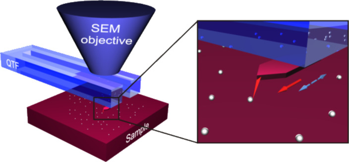
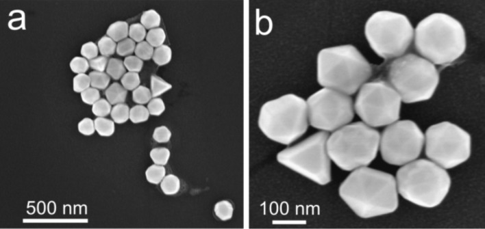
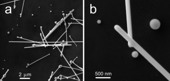
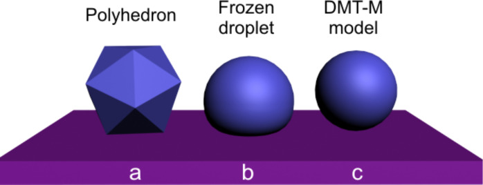
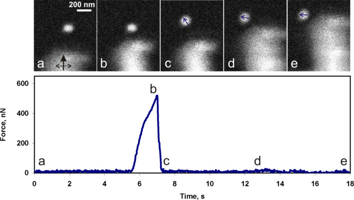
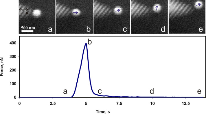
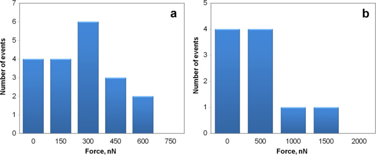
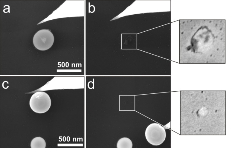
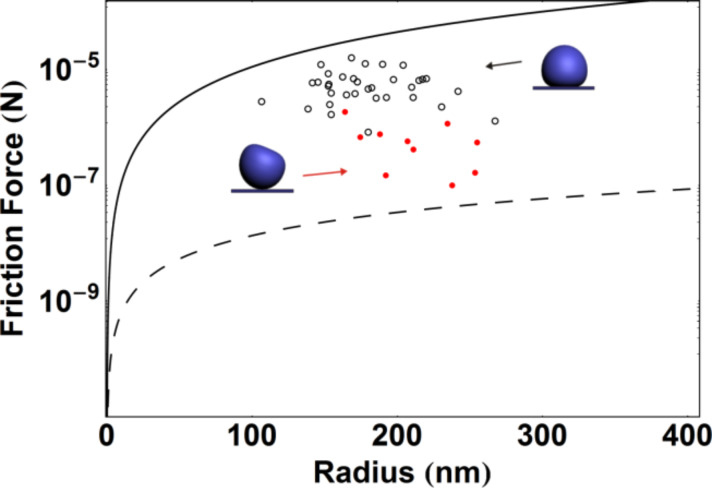
In this work polyhedron-like gold and sphere-like silver nanoparticles (NPs) were manipulated on an oxidized Si substrate to study the dependence of the static friction and the contact area on the particle geometry. Measurements were performed inside a scanning electron microscope (SEM) that was equipped with a high-precision XYZ-nanomanipulator. To register the occurring forces a quartz tuning fork (QTF) with a glued sharp probe was used. Contact areas and static friction forces were calculated by using different models and compared with the experimentally measured force. The effect of NP morphology on the nanoscale friction is discussed.

Keywords: contact mechanics; nanomanipulation; nanoparticles; nanotribology; scanning electron microscopy (SEM).


Figures

Figure 1
Figure 1
Schematics of the manipulation experiments inside an SEM. Solid arrow indicates the direction of the tip movement. Dashed arrow indicates QTF oscillation direction.
Figure 2
Figure 2
High resolution SEM images of Au NPs (150 nm) of different shape as deposited from a solution.
Figure 3
Figure 3
High resolution SEM images of Ag nanowires (diameter 120 nm) after pulsed laser annealing (a). Ag NPs of different size produced by laser annealing (b).
Figure 4
Figure 4
Different models for the estimation of the contact area: facet area of a polyhedron for Au NPs (a), frozen droplet for Ag NPs solidified on a substrate (b), DMT-M model for Ag NPs solidified without contact to a substrate (c).
Figure 5
Figure 5
SEM snapshots of the manipulation process of a Au NP by using a tungsten tip, and the corresponding force curve. The black solid arrow in image (a) indicates the movement direction of the tip and dashed arrow shows tip the oscillation direction. The small blue arrows indicate the NP displacement directions (b, c, d).
Figure 6
Figure 6
Snapshots of the manipulation of a Ag NP by using an AFM tip, and the corresponding force curve. The black solid arrow in image (a) indicates the tip movement direction and the dashed arrow shows the tip oscillation direction. The small blue arrows indicate NP displacement directions (b, c, d, e).
Figure 7
Figure 7
Distribution histogram of static friction force values that were experimentally measured for NPs of different shapes: polyhedron-like Au NPs (a) and for sphere-like Ag NPs (b), respectively.
Figure 8
Figure 8
High resolution SEM images of Ag NPs (no force recording during the displacement of the NPs). Traces indicating the contact area after the first (a,b) and the second (c,d) displacement of two different Ag NPs. The corresponding estimated static friction forces are ca. 6190 nN and about 930 nN, respectively.
Figure 9
Figure 9
The static friction force of Ag NPs on a Si wafer as a function of the radius of the NPs. The static friction force values experimentally measured by QTF (dots) and calculated from the diameters of visible traces left after the displacement of the particles (circles). The theoretical curves of friction as functions of the radius of the NPs according to the DMT-M model (dashed curve) and frozen droplet model (solid curve).

References

    1. Gnecco E, Meyer E, editors. Fundamentals of Friction and Wear. Berlin, Heidelberg, New York: Springer; 2007.
    1. Wang N, Rokhlin S I, Farson D F. Nanotechnology. 2008;19:415701. doi: 10.1088/0957-4484/19/41/415701. - DOI - PubMed
    1. Dietzel D, Ritter C, Mönninghoff T, Fuchs H, Schirmeisen A, Schwarz U D. Phys Rev Lett. 2008;101:125505. doi: 10.1103/PhysRevLett.101.125505. - DOI - PubMed
    1. Kim S, Ratchford D C, Li X. ACS Nano. 2009;3:2989–2994. doi: 10.1021/nn900606s. - DOI - PubMed
    1. Darwich S, Mougin K, Rao A, Gnecco E, Jayaraman S, Haidara D. Beilstein J Nanotechnol. 2011;2:85–98. doi: 10.3762/bjnano.2.10. - DOI - PMC - PubMed

LinkOut - more resources