. 2014 Dec;55(12):2917-23.
doi: 10.3109/10428194.2014.900762. Epub 2014 Apr 23.

A new model of LMP1-MYC interaction in B cell lymphoma

Affiliations

A new model of LMP1-MYC interaction in B cell lymphoma

Evelena P Ontiveros et al. Leuk Lymphoma. 2014 Dec.

Abstract

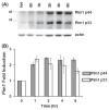
Epstein-Barr virus (EBV) is associated with aggressive B cell lymphomas (BCLs). Latent membrane protein 1 (LMP1) of EBV is an oncogenic protein required for EBV B cell transformation. However, LMP1 is a weak oncogene in mice. Mice expressing Myc inserted 5' of the Eμ enhancer (iMyc(Eμ)), mimicking the t(8;14) translocation of endemic Burkitt lymphoma, develop delayed onset BCLs. To investigate potential cooperation between LMP1 and oncogenic MYC, we produced mice expressing the LMP1 signaling domain via a hybrid CD40-LMP1 transgene (mCD40-LMP1), and the dysregulated MYC protein of aggressive EBV+ BCLs. mCD40-LMP1/iMyc(Eμ) mice trended toward earlier BCL onset. BCLs from mCD40-LMP1/iMyc(Eμ) mice expressed LMP1 and were transplantable into immunocompetent recipients. iMyc(Eμ) and mCD40-LMP1/iMyc(Eμ) mice developed BCLs with similar immunophenotypes. LMP1 signaling was intact in BCLs as shown by inducible interleukin-6. Additionally, LMP1 signaling to tumor cells induced the two isoforms of Pim1, a constitutively active prosurvival kinase implicated in lymphomagenesis.

Keywords: LMP1; MYC; lymphoma.


Conflict of interest statement

Potential conflict of interest: Disclosure forms provided by the authors are available with the full text of this article at www.informahealthcare.com/lal.

Figures

Figure 1
Figure 1
Lymphoma-free survival of mCD40–LMP1/iMyc mice. Kaplan–Meier survival analysis of mice of indicated genotypes. Median lymphoma-free survival for mCD40–LMP1/iMyc mice and iMyc mice was 227 and 271 days, respectively (p = 0.6953 as analyzed by two-sample Kolmogorov–Smirnov test). None of the mCD40–LMP1 or CD40−/− littermates developed lymphoma. All mice in the cohort were on a CD40−/− background.
Figure 2
Figure 2
Representative lymphoma from a mCD40–LMP1/iMyc transgenic mouse. (A) Lymphoblastic lymphoma histology. Splenic white pulp is expanded and red pulp infiltrated by neoplastic round cells. Neoplastic cells are round to polygonal with large nuclei containing 1–2 large nucleoli, often adhered to the nuclear membrane. The mitotic rate is high and macrophages containing apoptotic bodies are present. Hematoxylin and eosin (HE), magnification × 200, bar =100 μm. (Inset: HE, × 600.) (B) B220 staining. Neoplastic cells are B220 +. B220, magnification × 200, bar = 100 μm. (Inset: B220, × 600.) (C) LMP1 staining. LMP1 + tumor cells are effacing and infiltrating the kidney. LMP1, magnification × 200, bar = 100 μm. (Inset: LMP1, × 600.) All images were obtained with an Olympus BX51 microscope, attached Olympus DP72 camera and MicroSuite Pathology Software (Olympus). (D) Immunophenotyping of representative lymphoma. Representative lymphoma flow cytometry from a mCD40–LMP1/iMyc mouse. Black solid lines indicate antibody-specific staining and gray dashed lines indicate antibody isotype controls.
Figure 3
Figure 3
LMP1-induced IL-6 production in lymphoma cell lines derived from mCD40–LMP1/iMyc mice. Hal2G1 and Hal18 lymphoma cell lines were derived from separate mCD40–LMP1/iMyc mice and express the mCD40–LMP1 transgene but no native mouse CD40. Hal2G1 and Hal18 were stimulated with cells expressing CD154, which stimulates the mCD40–LMP1 protein to signal via the cytoplasmic LMP1 tail. The Hal17 lymphoma cell line was derived from an iMyc mouse, and does not express the mCD40–LMP1 transgene or endogenous mouse CD40. Triplicate replicates of each lymphoma cell line were cultured in BCM-10 alone (BCM), with control insect cells (control) or with insect cells expressing CD154 (CD154). Supernatants from 48 h time-points were collected and analyzed by mouse IL-6 ELISA. Data shown are representative of five independent experiments.
Figure 4
Figure 4
Pim1 expression in lymphoma cell lines derived from mCD40–LMP1/iMyc mice. (A) Inducible Pim1 expression. Hal2G1 lymphoma cells were stimulated with isotype control antibody (Iso) for 2 h or stimulated with agonistic anti-mouse CD40 antibody for the indicated times to induce the mCD40–LMP1 protein to signal via the cytoplasmic LMP1 tail. Cytoplasmic extracts were separated by SDS-PAGE and then analyzed by immunoblotting with an anti-Pim1 antibody that recognized both Pim1 p44 and p33 isoforms. As a loading control, blots were stripped and reprobed with anti-actin antibody. (B) Pim1 fold induction. Pim1 p44, Pim1 p33 and actin values were quantitated by densitometry. Pim1 densitometry values (arbitrary units [AUs]) for each isoform were divided by actin AUs for each lane. To calculate fold induction, Pim1 AU/actin AU values were normalized to the 0 h (Pim1 AU/actin AU) value which was arbitrarily normalized to 1. Similar results were seen in two other cell lines (Hal18 and hCD40–LMP1 CH12.LX cells), which also express an inducible LMP1 transgene (data not shown). Data shown are representative of six independent experiments.

References

    1. Graham JP, Arcipowski KM, Bishop GA. Differential B-lymphocyte regulation by CD40 and its viral mimic, latent membrane protein 1. Immunol Rev. 2010;237:226–248. - PubMed
    1. Heslop HE. Biology and treatment of Epstein-Barr virus-associated non-Hodgkin lymphomas. Hematol Am Soc Hematol Educ Prog. 2005:260–266. - PubMed
    1. Stunz LL, Busch LK, Munroe ME, et al. Expression of the cytoplasmic tail of LMP1 in mice induces hyperactivation of B lymphocytes and disordered lymphoid architecture. Immunity. 2004;21:255–266. - PubMed
    1. Rastelli J, Homig-Holzel C, Seagal J, et al. LMP1 signaling can replace CD40 signaling in B cells in vivo and has unique features of inducing class-switch recombination to IgG1. Blood. 2008;111:1448–1455. - PubMed
    1. Kulwichit W, Edwards RH, Davenport EM, et al. Expression of the Epstein-Barr virus latent membrane protein 1 induces B cell lymphoma in transgenic mice. Proc Natl Acad Sci USA. 1998;95:11963–11968. - PMC - PubMed

Publication types

MeSH terms

Substances

LinkOut - more resources