. 2014 Jun;99(6):E1022-30.
doi: 10.1210/jc.2014-1118. Epub 2014 Feb 28.

Noninvasive prenatal diagnosis of congenital adrenal hyperplasia using cell-free fetal DNA in maternal plasma

Affiliations

Noninvasive prenatal diagnosis of congenital adrenal hyperplasia using cell-free fetal DNA in maternal plasma

Maria I New et al. J Clin Endocrinol Metab. 2014 Jun.

Abstract

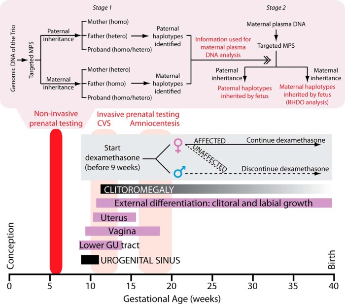
Context: Congenital adrenal hyperplasia (CAH) is an autosomal recessive condition that arises from mutations in CYP21A2 gene, which encodes for the steroidogenic enzyme 21-hydroxylase. To prevent genital ambiguity in affected female fetuses, prenatal treatment with dexamethasone must begin on or before gestational week 9. Currently used chorionic villus sampling and amniocentesis provide genetic results at approximately 14 weeks of gestation at the earliest. This means that mothers who want to undergo prenatal dexamethasone treatment will be unnecessarily treating seven of eight fetuses (males and three of four unaffected females), emphasizing the desirability of earlier genetic diagnosis in utero.

Objective: The objective of the study was to develop a noninvasive method for early prenatal diagnosis of fetuses at risk for CAH.

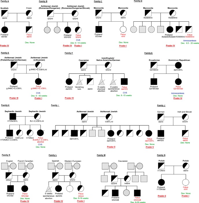
Patients: Fourteen families, each with a proband affected by phenotypically classical CAH, were recruited.

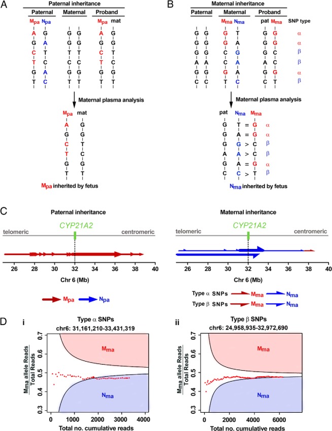
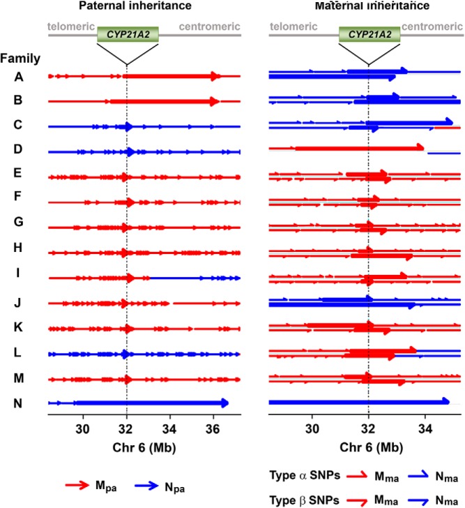
Design: Cell-free fetal DNA was obtained from 3.6 mL of maternal plasma. Using hybridization probes designed to capture a 6-Mb region flanking CYP21A2, targeted massively parallel sequencing (MPS) was performed to analyze genomic DNA samples from parents and proband to determine parental haplotypes. Plasma DNA from pregnant mothers also underwent targeted MPS to deduce fetal inheritance of parental haplotypes.

Results: In all 14 families, the fetal CAH status was correctly deduced by targeted MPS of DNA in maternal plasma, as early as 5 weeks 6 days of gestation.

Conclusions: MPS on 3.6 mL plasma from pregnant mothers could potentially provide the diagnosis of CAH, noninvasively, before the ninth week of gestation. Only affected female fetuses will thus be treated. Our strategy represents a generic approach for noninvasive prenatal testing for an array of autosomal recessive disorders.


Figures

Figure 1.
Figure 1.
Conventional prenatal management and targeted MPS for noninvasive detection of CAH. Temporal relationship between normal genital organ development, detection of CAH mutations by CVS or amniocentesis, and initiation of therapy with dexamethasone are shown. Targeted MPS should provide an early diagnosis before 9 weeks of gestation. In stage 1, genomic DNA samples of the mother, father, and proband (trio) were subjected to targeted MPS of the CYP21A2 region. For paternal inheritance, SNPs that were homozygous (homo) in the mother and heterozygous (hetero) in the father were used. Paternal haplotypes inherited by the proband and absent in the proband were thus identified. For maternal inheritance, SNPs that were heterozygous in the mother and homozygous in the father were used. Maternal haplotypes (linked or not linked with the proband's mutation) were determined. Stage 2 targeted MPS of the CYP21A2 region on plasma cell-free DNA of the pregnant mother. Detection of the paternal-specific alleles in maternal plasma revealed the inheritance of either the paternal haplotype linked or not linked with the proband's mutation from the father. Maternal inheritance was determined by the RHDO analysis (see Supplemental Methods and Reference for details).
Figure 2.
Figure 2.
Pedigree for 14 families with CAH that underwent massively parallel sequencing and haplotype analysis. The parents and proband constituted the trio. Genotype, ethinicity, Prader score (for females), and whether the mother received prenatal dexamethasone are shown. Please read in conjunction with Table 1.
Figure 3.
Figure 3.
Fetal inheritance of parental mutations in family A. A, For paternal inheritance, SNPs that were homozygous in the mother and heterozygous in the father were selected. The paternal-specific SNP alleles transmitted to the proband belonged to the haplotype linked with the proband's mutation (Mpa, red). The paternal-specific SNP alleles not present in the proband belonged to the haplotype not linked with the proband's mutation (Npa, blue). B, For maternal inheritance, SNPs that were heterozygous in the mother and homozygous in the father were classified into type α- or type β-SNPs. For type α-SNPs, the paternal alleles were the same as those on Mma (red). The equal representation of Mma and Nma alleles in the maternal plasma indicated that the fetus had inherited Nma from the mother. For type β-SNPs, the paternal alleles were the same as those of Nma (blue). The overrepresentation of Nma alleles in the maternal plasma also indicated that the fetus had inherited Nma from the mother. C, Parental haplotype blocks are denoted by arrows along the entire targeted region. The tail and tip of an arrow denote the start and end, respectively, of a RHDO block, established analytically from the telomeric to the centromeric end. Left, Paternal inheritance analysis resulted in 14 paternal haplotype blocks, represented by red arrows, all indicating the inheritance of Mpa. Prenatal determination of the fetal inheritance status was made according to the haplotype block spanning the CYP21A2 gene, indicated with a thick red arrow. Right, Maternal inheritance analysis resulted in six and one SPRT classifications using type α- and type β-SNPs, respectively. The haplotypes established by the type α- and β-SNPs are represented by upper and lower half-arrows, respectively. The fetus had inherited Nma from the mother by both type α- and β-SNPs. The SPRT classification near the centromeric end indicated that a recombination event had occurred. Grey line, targeted region spanning chromosome 6: 24,954,958–38,933,706. Dotted line and green box, CYP21A2 gene on chromosome 6: 32,006,093–32,009,447. D, RHDO analysis showing SPRT classification process using type α-SNPs on chromosome 6: 31,161,210–33,431,319 (i) or type β-SNPs on chromosome 6: 24,958,935–32,972,690 (ii). For details, please refer to Supplemental Methods.
Figure 4.
Figure 4.
Fetal haplotype analysis in families A to N. A haplotype block is indicated by an arrow. The tail and tip of an arrow denotes the start and end, respectively, of a haplotype block, established analytically from the telomeric to the centromeric end. Left, Fetal inheritance of the paternal haplotype was determined by the presence of paternal-specific SNP alleles in maternal plasma DNA samples. The haplotype block spanning the CYP21A2 gene is denoted by a thick arrow in each family. Right, Fetal inheritance of the maternal haplotype was determined by RHDO analysis. Haplotype blocks within chromosome 6, 28–35 Mb, are shown. Upper and lower half-arrows denote the SPRT classification made by type α- or type β-SNPs, respectively. The SPRT classification spanning the CYP21A2 gene region, denoted by a thick half-arrow, indicates the maternal haplotype inherited by the fetus. Notably, the mother and father of families G and H, respectively, were heterozygous for two CYP21A2 gene mutations themselves. Targeted MPS analysis indicated that the fetuses in both families had inherited the haplotypes linked to the proband's mutations from the affected parents. The dotted lines indicate the location of the CYP21A2 gene (green box). The haplotype blocks of several samples are partially shown, as indicated by a line without an arrow tip toward the centromeric end. In family H, a recombination event in the maternal allele, which occurs outside of the chromosomal region displayed above, is not shown.

References

    1. Pang SY, Wallace MA, Hofman L, et al. Worldwide experience in newborn screening for classical congenital adrenal hyperplasia due to 21-hydroxylase deficiency. Pediatrics. 1988;81:866–874 - PubMed
    1. New MI, Abraham M, Gonzalez B, et al. Genotype-phenotype correlation in 1,507 families with congenital adrenal hyperplasia owing to 21-hydroxylase deficiency. Proc Natl Acad Sci USA. 2013;110:2611–2616 - PMC - PubMed
    1. New MI, Lekarev O, Mancenido D, Parsa A, Yuen T. Congenital adrenal hyperplasia owing to 21-hydroxylase deficiency. In: New MI, Lekarev O, Parsa A, Yuen T, O'Malley BW, Hammer GD, eds. Genetic Steroid Disorders. London: Elsevier; 2014:29–51
    1. Haider S, Islam B, D'Atri V, et al. Structure-phenotype correlations of human CYP21A2 mutations in congenital adrenal hyperplasia. Proc Natl Acad Sci USA. 2013;110:2605–2610 - PMC - PubMed
    1. New MI, Abraham M, Yuen T, Lekarev O. An update on prenatal diagnosis and treatment of congenital adrenal hyperplasia. Semin Reprod Med. 2012;30:396–399 - PubMed

Publication types