Review
. 2014:83:129-57.
doi: 10.1146/annurev-biochem-060713-035314. Epub 2014 Mar 6.

Demystifying heparan sulfate-protein interactions

Affiliations
Review

Demystifying heparan sulfate-protein interactions

Ding Xu et al. Annu Rev Biochem. 2014.

Abstract

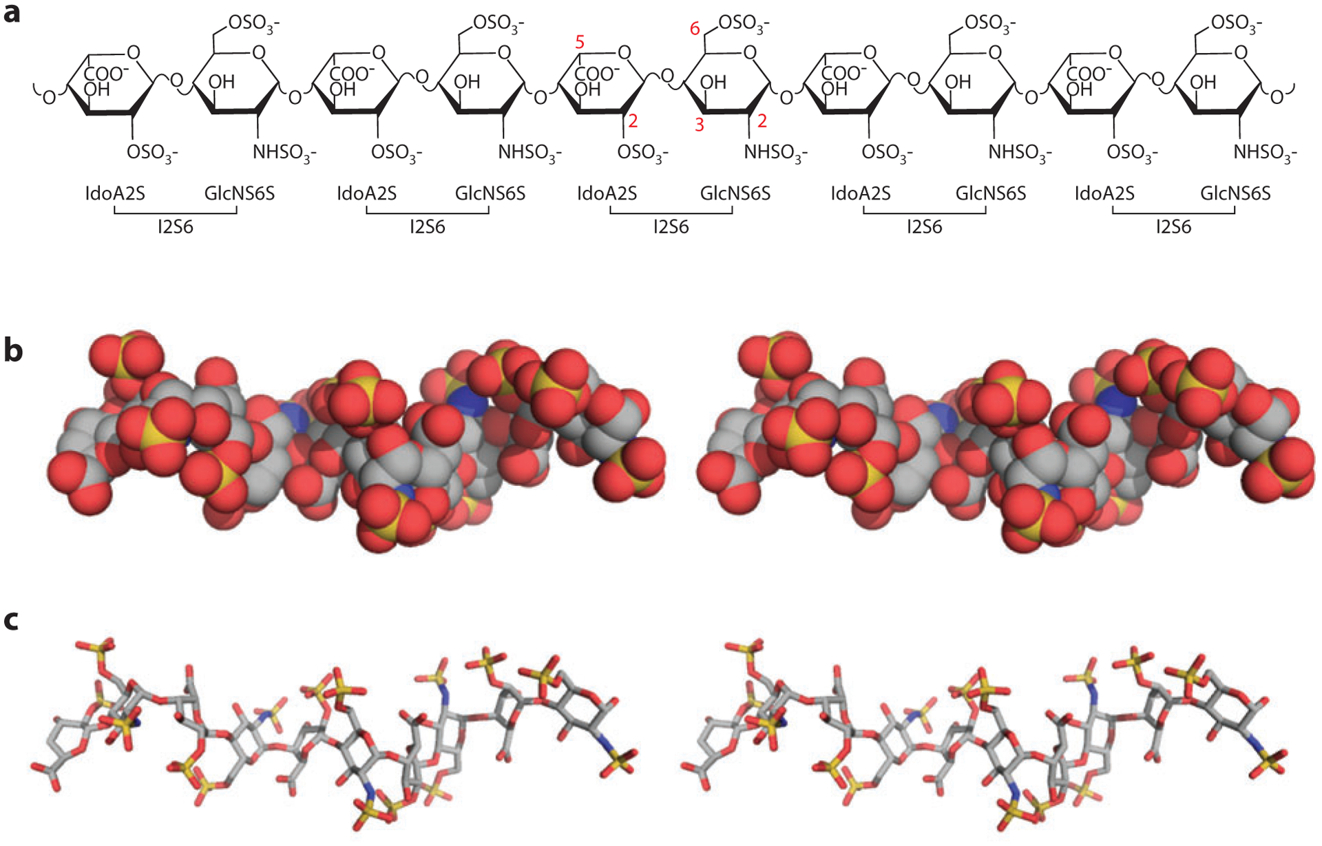
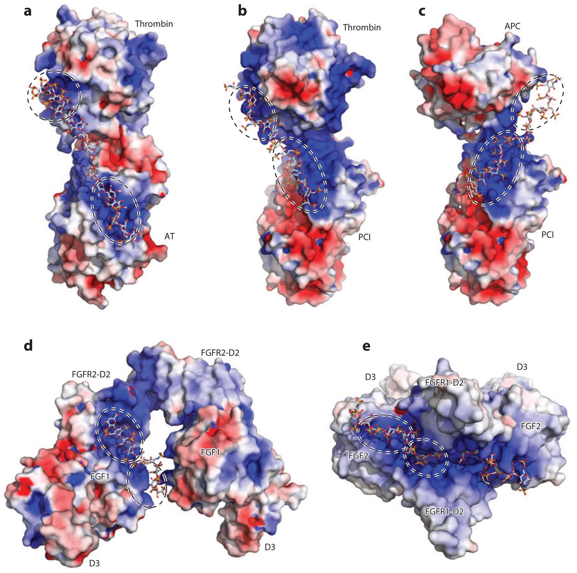
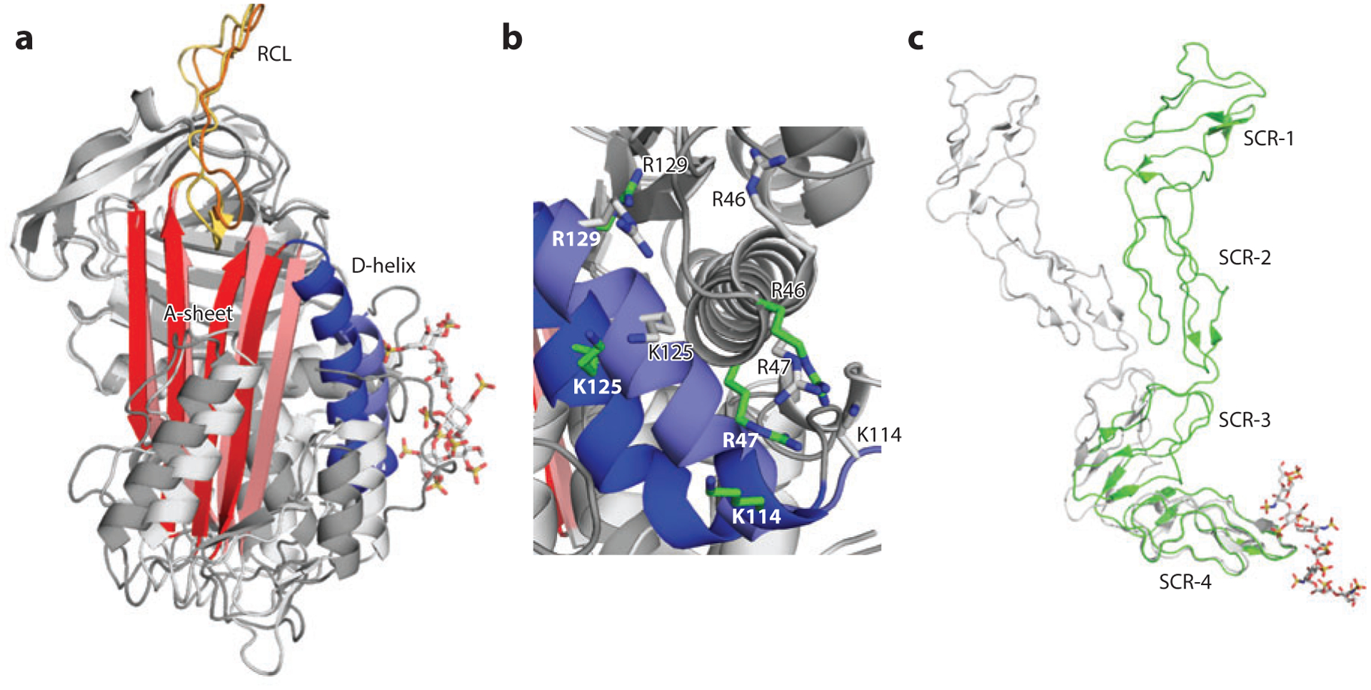
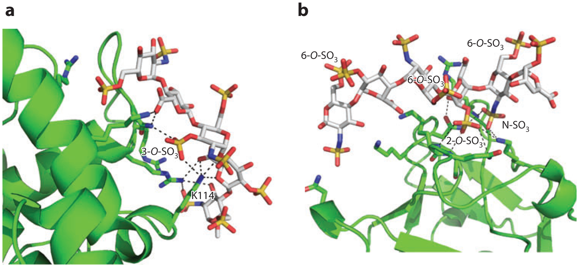
Numerous proteins, including cytokines and chemokines, enzymes and enzyme inhibitors, extracellular matrix proteins, and membrane receptors, bind heparin. Although they are traditionally classified as heparin-binding proteins, under normal physiological conditions these proteins actually interact with the heparan sulfate chains of one or more membrane or extracellular proteoglycans. Thus, they are more appropriately classified as heparan sulfate-binding proteins (HSBPs). This review provides an overview of the various modes of interaction between heparan sulfate and HSBPs, emphasizing biochemical and structural insights that improve our understanding of the many biological functions of heparan sulfate.

Keywords: glycan–protein interaction; glycosaminoglycan; heparan sulfate–binding domain; heparin-binding protein; oligomerization; proteoglycan.


Figures

Figure 1
Figure 1
Structure of heparan sulfate. (a) Chemical structure of a heparin-derived decasaccharide. Carbon numbers of the modification sites are indicated in red. (b) Stereo view of a heparin-derived decasaccharide in space-filling representation, based on Protein Data Bank identifier 1E0O, with carbon in gray, oxygen in red, nitrogen in blue, and sulfate in yellow. (c) Stereo view of the same decasaccharide in stick representation. Note the helical nature of the chain and the alternating clusters of three sulfates groups on each side of the sugar backbone. Abbreviations: GlcNS, N-sulfoglucosamine; IdoA, L-iduronic acid.
Figure 2
Figure 2
Heparan sulfate (HS)–induced HS-binding protein oligomerization. In the three-dimensional drawings, one monomer is in green and one in pink; in the surface representations, positive electrostatic potential is in blue and negative electrostatic potential is in red. For the oligosaccharide, carbon is gray, oxygen red, nitrogen blue, and sulfate yellow. (a) Structure of FGF1 (fibroblast growth factor 1) dimer and bound oligosaccharide [Protein Data Bank (PDB) identifier 1AXM]. (b) Structure of the dimeric V-C1 domains of RAGE (receptor for advanced glycation end products) (PDB 4IM8). The dodecasaccharide is manually modeled into the structure on the basis of the observed partial electron density. (c) Structure of the dimeric E2 domain of amyloid precursor–like protein 1 (APLP-1) and bound oligosaccharide (PDB 3QMK). (d) Structure of dimeric interleukin-8 (PDB 2IL8) and a modeled oligosaccharide (degree of polymerization: 20). (e) Structure of dimeric CXCL12 (PDB 2NWG) and a modeled decasaccharide.
Figure 3
Figure 3
Heparan sulfate (HS) acts as a molecular scaffold. In all structures, proteins are shown in surface electrostatic potential, and the oligosaccharides are shown in stick representation. Each HS-binding site is enclosed in a dashed circle. (a) Cocrystal structure of thrombin, antithrombin (AT), and a hexadescasaccharide heparin mimic [Protein Data Bank (PDB) identifier 1TB6]. (b) Cocrystal structure of thrombin, protein C inhibitor (PCI), and a heparin-derived tetradecasaccharide. Because the tetradecasaccharide cannot be fully resolved in the cocrystal structure except for two sugar residues, it is manually modeled here (PDB 3B9F). (c) A model of the ternary complex of activated protein C (APC), PCI, and tetradecasaccharide based on PDB 3B9F and biochemical evidence (92). (d) Cocrystal structure of fibroblast growth factor 1 (FGF1), FGF receptor 2 (FGFR2, D2 and D3 domains), and heparin-derived decasaccharide (PDB 1E0O). (e) Cocrystal structure of FGF2, FGFR1 (D2 and D3 domains), and heparin-derived decasaccharide (PDB 1FQ9).
Figure 4
Figure 4
Heparan sulfate (HS) acts as an allosteric regulator. (a) Overlay of native antithrombin [Protein Data Bank (PDB) identifier 1E04) (light gray) and pentasaccharide-bound antithrombin (PDB 1AZX) (dark gray). The important structural elements are the D-helix in purple (native) and blue (bound), the A-sheet in pink (native) and red (bound), and the reactive central loop (RCL) in yellow (native) and orange (bound). (b) Rearrangement of HS-binding residues following pentasaccharide binding. For clarity, the pentasaccharide is not displayed. The native conformation of HS-binding residues is shown as gray sticks, and the pentasaccharide-bound conformation of HS-binding residues is shown as green sticks. (c) Overlay of native Vaccinia virus complement control protein (VCP) (PDB 1G40) (gray) and decasaccharide-bound VCP (PDB 1RID) (green). Abbreviation: SCR, short consensus repeat.
Figure 5
Figure 5
Structural basis of modification-specific heparan sulfate (HS)–HS-binding protein (HSBP) interactions. (a) Pentasaccharide–antithrombin (AT) interaction [Protein Data Bank (PDB) identifier 2GD4]. HS-binding residues of AT are shown as green sticks, and the pentasaccharide is shown as gray sticks with oxygen in red, nitrogen in blue, and sulfate in yellow. The ionic and nonionic interactions stabilized by 3-O-sulfate and K114 (both directly and indirectly) are shown as dashed lines. (b) Hexasaccharide–fibroblast growth factor 2 (FGF2) interaction (PDB 1BFC). HS-binding residues and hexasaccharide are colored as in panel a. The ionic and nonionic interactions that involve the critical disaccharide—IdoA2S-GlcNS6S—are shown as dashed lines.

References

    1. Guerardel Y, Czeszak X, Sumanovski LT, Karamanos Y, Popescu O, et al. 2004. Molecular fingerprinting of carbohydrate structure phenotypes of three Porifera proteoglycan-like glyconectins. J. Biol. Chem 279:15591–603 - PubMed
    1. Yamada S, Morimoto H, Fujisawa T, Sugahara K. 2007. Glycosaminoglycans in Hydra magnipapillata (Hydrozoa, Cnidaria): demonstration of chondroitin in the developing nematocyst, sting organelle, and structural characterization of glycosaminoglycans. Glycobiology 17:886–94 - PubMed
    1. Lawrence R, Olson SK, Steele RE, Wang L, Warrior R, et al. 2008. Evolutionary differences in glycosaminoglycan fine structure detected by quantitative glycan reductive isotope labeling. J. Biol. Chem 283:33674–84 - PMC - PubMed
    1. Ori A, Wilkinson MC, Fernig DG. 2011. A systems biology approach for the investigation of the heparin/heparan sulfate interactome. J. Biol. Chem 286:19892–904 - PMC - PubMed
    1. Bishop JR, Schuksz M, Esko JD. 2007. Heparan sulphate proteoglycans fine-tune mammalian physiology. Nature 446:1030–37 - PubMed

Publication types