Review
. 2014:83:441-66.
doi: 10.1146/annurev-biochem-060713-035524. Epub 2014 Mar 5.

Hierarchy of RNA functional dynamics

Affiliations
Review

Hierarchy of RNA functional dynamics

Anthony M Mustoe et al. Annu Rev Biochem. 2014.

Abstract

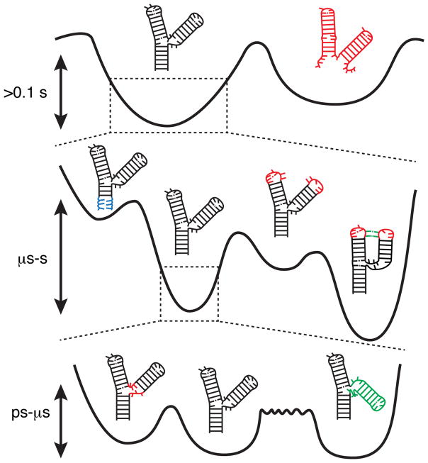
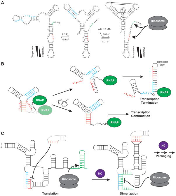
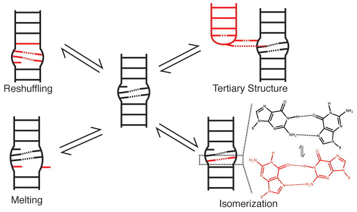
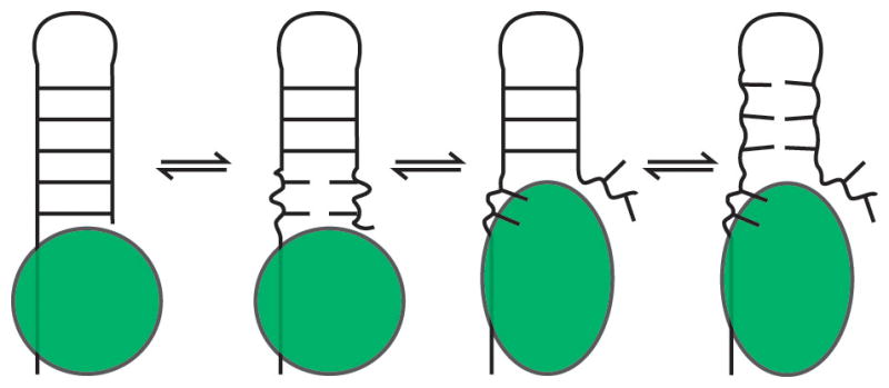
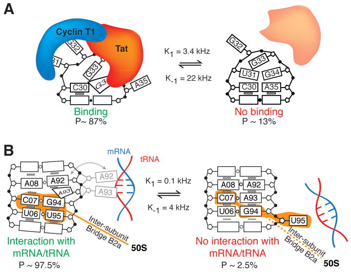
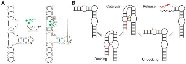
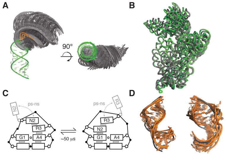
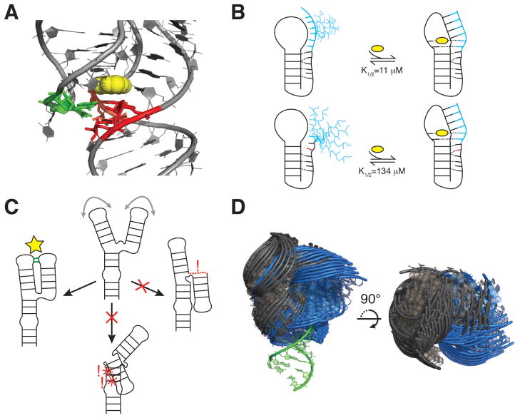
RNA dynamics play a fundamental role in many cellular functions. However, there is no general framework to describe these complex processes, which typically consist of many structural maneuvers that occur over timescales ranging from picoseconds to seconds. Here, we classify RNA dynamics into distinct modes representing transitions between basins on a hierarchical free-energy landscape. These transitions include large-scale secondary-structural transitions at >0.1-s timescales, base-pair/tertiary dynamics at microsecond-to-millisecond timescales, stacking dynamics at timescales ranging from nanoseconds to microseconds, and other "jittering" motions at timescales ranging from picoseconds to nanoseconds. We review various modes within these three different tiers, the different mechanisms by which they are used to regulate function, and how they can be coupled together to achieve greater functional complexity.

Keywords: RNA catalysis; RNA flexibility; molecular adaptation; regulatory RNA; riboswitches.


Conflict of interest statement

DISCLOSURE STATEMENT

H.M.A. is an advisor to and holds an ownership interest in Nymirum Inc., which is an RNA-based drug discovery company. The research reported in this article was performed by the University of Michigan faculty and students and was funded by an NIH contract to H.M.A.

Figures

Figure 1
Figure 1
The different tiers of RNA dynamics. At the lowest level of the hierarchy are secondary structure dynamics, which define broad free energy basins with high separating barriers. Within each secondary structure are smaller alternative base pairing arrangements that define Tier 1 dynamics. These include base pair melting (blue, left), reshuffling (middle right, red), and tertiary pairing (green). Each local pairing basin in turn defines a limited set of 3D conformations, transitions between which comprise Tier 2 dynamics. These dynamics include loop dynamics (left, red) and inter-helical dynamics (right, green). Although inter-helical and loop-dynamics have similar barrier heights, due to the larger number of involved coordinates inter-helical dynamics typically proceed more slowly (long rough separating barrier).
Figure 2
Figure 2
(A) Three state secondary structure equilibrium of the add adenine riboswitch. In the adenine-bound conformation both the start codon (green) and ribosome binding site (red) are exposed, upregulating translation. The temperature dependence of the apo-state secondary structure equilibrium offsets the increased ligand affinity of the binding-competent conformation at low temperature (30). (B) Example of a transcriptional acting adenine riboswitch. Ligand binding stabilizes a transient secondary structure, sequestering residues that would otherwise pair with downstream transcribed sequences to form the thermodynamically favored terminator stem. (C) The HIV-1 5’ leader couples exposure of the start codon of the downstream-encoded gag protein to sequestration of the dimerization initiation site (DIS; red), promoting translation while inhibiting dimerization (left). In a process promoted by the nucleocapsid chaperone protein (NC; purple), the leader undergoes a secondary structure switch that exposes the DIS and sequesters the start codon, attenuating translation and promoting dimerization, which initiates genome packaging (right) (40).
Figure 3
Figure 3
Modes of Tier 1 dynamics.
Figure 4
Figure 4
Example of an RNA chaperone. The bound chaperone destabilizes the neighboring RNA helix, promoting melting dynamics, and then binds the exposed nucleotides. The other strand is released and can interact with other RNAs, and the remainder of the helix is also destabilized.
Figure 5
Figure 5
Functions of base-pair reshuffling dynamics. (A) In the HIV-1 TAR apical loop, the minor CS sequesters residues involved in HIV Tat and Cyclin T1 binding during transcriptional activation (54). (B) In the major CS of the ribosomal A-site, A92 and A93 are free to interact with and stabilize cognate mRNA/tRNA minihelices during decoding, indicated by the gray dashed arrow and alternative A92/A93 conformation. The minor CS sequesters these residues, inhibiting decoding and also disrupting the B2a inter-subunit bridge (54).
Figure 6
Figure 6
(A) Coupling of base-reshuffling and tertiary dynamics in the P5abc domain of the Tetrahymena thermophila group I ribozyme. Upon binding of two Mg2+ ions (184), the P5c stem (colored) undergoes a 1-nt register shift, releasing U168 to participate in a long-range pair (right, boxed). Additional tertiary interactions, which are not shown, are also formed upon folding. NMR studies observed the two conformations to be in slow exchange (87), with agreement from recent stopped flow experiments (88). Dashed lines indicate non-canonical pairs. (B) Enzymatic cycle of the hairpin ribozyme (91).
Figure 7
Figure 7
Modes and functions of Tier 2 jittering dynamics. (A) View of 50 most probable inter-helical conformations for a 3-nt two-helix bulge junction with the lower stem superimposed (green). Most probable conformations were obtained from coarse-grained model simulations that include only steric and connectivity forces (178). Bulge residues were included in coarse-grained modeling but not shown in the figure, instead drawn as orange lines highlighting the possible paths of the bulge. (B) Superposition of classical (green; PDB ID 2WDG) and ratcheted EF-G bound 16S rRNA conformations (grey; PDB IDB 4JUW), highlighting the large inter-helical dynamics associated with ribosomal translocation. The rRNAs were superimposed using residues 1410–1430 and 1470–1490 of H44, with H44 facing the page. (C) Dynamics of the GNRA tetraloop observed by fluorescence spectroscopy (159). Exchange timescales correspond to rates measured by base relaxation (55) and sugar carbon NMR relaxation dispersion experiments (158). (D) Superposition of ligand bound HIV-1 TAR structures (grey) with five conformers from a high-resolution NMR-MD ensemble that have the lowest heavy-atom RMSD to the ligand-bound structures (orange) (123). Left, PDB ID 1LVJ. Right, PDB ID 1UTS.
Figure 8
Figure 8
Interdependencies of CS across Tiers. (A) Aptamer domain of the add adenine riboswitch in complex with adenine (yellow) (PDB ID 1Y26). P1 stem base pairs shown to be unstable in the absence of ligand are shown in red (30), with J2/3 residues that provide stabilizing A-minor interactions shown in green. (B) Stacking interactions limit loop dynamics and pre-organize the 3’ tail for ligand binding and pseudoknot folding in the wild-type Bsu preQ1 riboswitch aptamer (top). An A-to-C mutation distal from the ligand binding pocket disrupts stacking, increasing dynamics and reducing ligand/riboswitch affinity (bottom) (176). The preQ1 ligand, 3’ tail, and mutation are shown in yellow, blue, and red, respectively. (C) Topological constraints preclude a three-way junction from forming two of three possible tertiary interactions. Right, the interaction is precluded due to connectivity. Bottom, the interaction is precluded due to sterics. (D) View of 50 most probable inter-helical conformations for two 1-nt bulge junctions with lower stem superimposed (green). The bulge of the blue junction is located two base-pairs below that of the grey junction. Most probable conformations were obtained from coarse-grained model simulations that include only steric and connectivity forces (178).

References

    1. Kruger K, Grabowski PJ, Zaug AJ, Sands J, Gottschling DE, Cech TR. Self-splicing RNA: autoexcision and autocyclization of the ribosomal RNA intervening sequence of Tetrahymena. Cell. 1982;31:147–57. - PubMed
    1. Guerrier-Takada C, Gardiner K, Marsh T, Pace N, Altman S. The RNA moiety of ribonuclease P is the catalytic subunit of the enzyme. Cell. 1983;35:849–57. - PubMed
    1. Serganov A, Patel DJ. Metabolite recognition principles and molecular mechanisms underlying riboswitch function. Annu Rev Biophys. 2012;41:343–70. - PMC - PubMed
    1. Reiter NJ, Chan CW, Mondragon A. Emerging structural themes in large RNA molecules. Curr Opin Struct Biol. 2011;21:319–26. - PMC - PubMed
    1. Voorhees RM, Ramakrishnan V. Structural basis of the translational elongation cycle. Annu Rev Biochem. 2013;82:203–36. - PubMed

Publication types