. 2014 Mar 7:14:92.
doi: 10.1186/1472-6882-14-92.

Electroacupuncture-like stimulation at Baihui and Dazhui acupoints exerts neuroprotective effects through activation of the brain-derived neurotrophic factor-mediated MEK1/2/ERK1/2/p90RSK/bad signaling pathway in mild transient focal cerebral ischemia in rats

Affiliations

Electroacupuncture-like stimulation at Baihui and Dazhui acupoints exerts neuroprotective effects through activation of the brain-derived neurotrophic factor-mediated MEK1/2/ERK1/2/p90RSK/bad signaling pathway in mild transient focal cerebral ischemia in rats

Chin Yi Cheng et al. BMC Complement Altern Med. .

Abstract

Background: This study was designed to evaluate the effects of electroacupuncture-like stimulation at Baihui (GV20) and Dazhui (GV14) acupoints (EA at acupoints) following mild cerebral ischemia-reperfusion (I/R) injury. Furthermore, we investigated whether brain-derived neurotrophic factor (BDNF)-mediated activation of extracellular signal-regulated kinase (ERK)1/2 signaling pathway is involved in the neuroprotection induced by EA at acupoints.

Methods: Rats were subjected to middle cerebral artery occlusion (MCAo) for 15 min followed by reperfusion for 3 d. EA at acupoints was applied 1 d postreperfusion then once daily for 2 consecutive days.

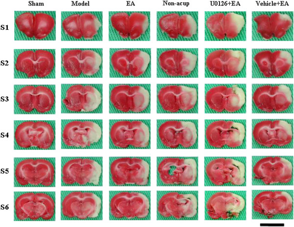
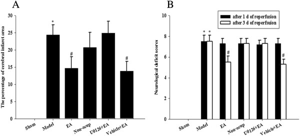
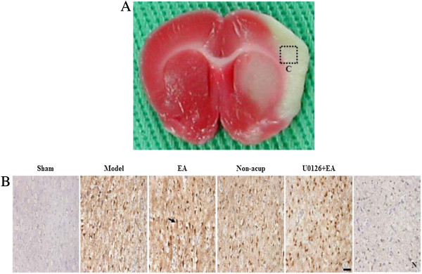
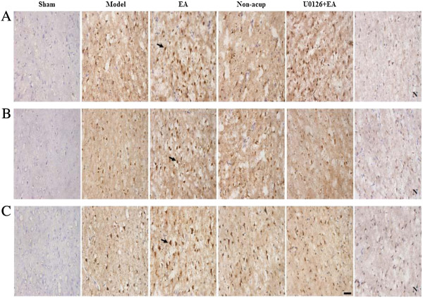
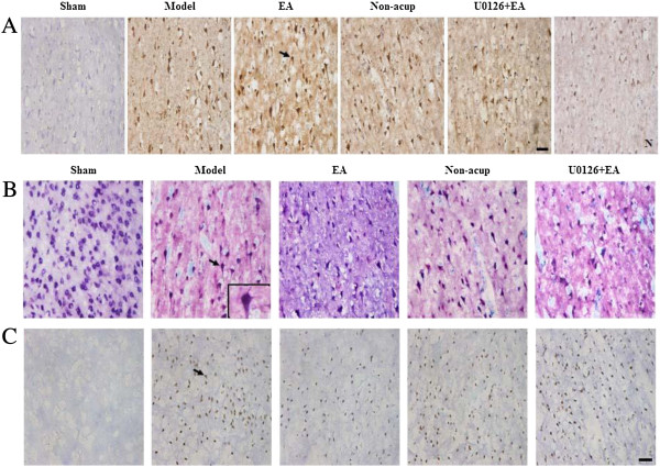
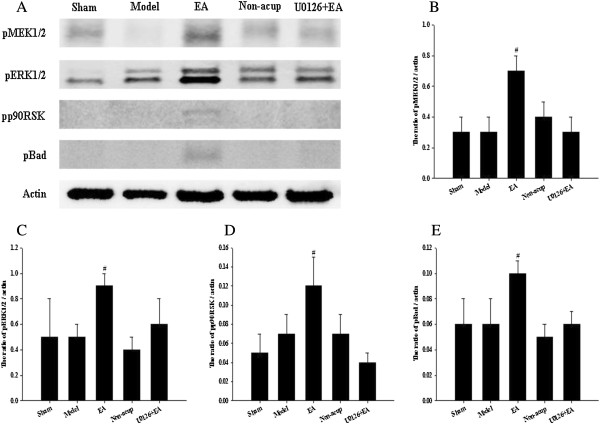
Results: Following the application of EA at acupoints, initiated 1 d postreperfusion, we observed significant reductions in the cerebral infarct area, neurological deficit scores, active caspase-3 protein expression, and apoptosis in the ischemic cortex after 3 d of reperfusion. We also observed markedly upregulated BDNF, phospho-Raf-1 (pRaf-1), phospho-MEK1/2 (pMEK1/2), phospho-ERK1/2 (pERK1/2), phospho-90 kDa ribosomal S6 kinase (pp90RSK), and phospho-Bad (pBad) expression, and restored neuronal nuclear antigen (NeuN) expression. Pretreatment with the MEK1/2 inhibitor U0126 abrogated the effects of EA at acupoints on cerebral infarct size, neurological deficits, active caspase-3 protein, and apoptosis in the ischemic cortex after 3 d of reperfusion. Pretreatment with U0126 also abrogated the effects of EA at acupoints on pMEK1/2, pERK1/2, pp90RSK, pBad, and NeuN expression, but did not influence BDNF and pRaf-1 expression.

Conclusion: Overall, our study results indicated that EA at acupoints, initiated 1 d postreperfusion, upregulates BDNF expression to provide BDNF-mediated neuroprotection against caspase-3-dependent neuronal apoptosis through activation of the Raf-1/MEK1/2/ERK1/2/p90RSK/Bad signaling cascade after 3 d of reperfusion in mild MCAo.


Figures

Figure 1
Figure 1
Focal cerebral infarct areas (S1 to S6) among the experimental groups after 15 min of ischemia followed by 3 d of reperfusion (n = 4 to 6). 2,3,5-Triphenyltetrazolium chloride staining showed the infarct area as white and the noninfarct area as red. Sham, sham group; Model, model group; EA, EA group; Non-acup, non-acup group; U0126 + EA, U0126 + EA group; Vehicle + EA, vehicle + EA group. Scale bar = 1 cm.
Figure 2
Figure 2
Effects of EA at acupoints on cerebral infarct and neurological status. (A) The percentage of cerebral infarct areas among the sham, model, EA, non-acup, U0126 + EA, and vehicle + EA groups were measured after 3 d of reperfusion. (B) The neurological deficit scores among the sham, model, EA, non-acup, U0126 + EA, and vehicle + EA groups were measured after 1 d and 3 d of reperfusion. Data are presented as mean ± SD. *P < 0.05 compared with the sham group; #P < 0.05 compared with the model group.
Figure 3
Figure 3
Effect of EA at acupoints on BDNF expression in the ischemic cortex. (A) Representative photograph showed a brain coronal section (TTC stain) from posterior bregma 0.92 mm. The dotted line square indicates the area of evaluation of immunopositive cells. C, the ischemic area of the cortex. Dotted line square = 1 mm2. (B) Representative photographs showed BDNF expression in the ischemic cortex of the sham, model, EA, non-acup, and U0126 + EA groups after 3 d of reperfusion. N, negative control stain. Arrow indicates a BDNF-positive cell. Scale bar = 50 μm.
Figure 4
Figure 4
Effects of EA at acupoints on pRaf-1, pMEK1/2, and pERK1/2 expression. (A) Representative photographs showed pRaf-1, (B) pMEK1/2, and (C) pERK1/2 expression in the ischemic cortex of the sham, model, EA, non-acup, and U0126 + EA groups after 3 d of reperfusion. N, negative control stain. Arrows indicate immunopositive cells. Scale bar = 50 μm.
Figure 5
Figure 5
Effects of EA at acupoints on pp90RSK, active caspase-3-NeuN, and TUNEL expression. (A) Representative photographs showed pp90RSK expression, (B) active caspase-3 (red) colocalizing with NeuN (blue), and (C) TUNEL-positive cells in the ischemic cortex of the sham, model, EA, non-acup, and U0126 + EA groups after 3 d of reperfusion. N, negative control stain. Arrows in (A) and (C) indicate pp90RSK- and TUNEL-positive cells, respectively. Arrow in (B) indicates active caspase-3-NeuN double-labeled cells (purple), shown at higher magnification in the bottom right panel. Scale bar = 50 μm.
Figure 6
Figure 6
Effects of EA at acupoints on cytosolic expression of pMEK1/2, pERK1/2, pp90RSK, and pBad. (A) Representative western blot images showed the cytosolic expression of pMEK1/2, pERK1/2, pp90RSK, and pBad in the ischemic cortex in the sham, model, EA, non-acup, and U0126 + EA groups after 3 d of reperfusion. Actin was used as an internal control. The relative cytosolic expression of (B) pMEK1/2, (C) pERK1/2, (D) pp90RSK, and (E) pBad (n = 4) was assessed in the ischemic cortex in the sham, model, EA, non-acup, and U0126 + EA groups. Data are presented as mean ± SD. #P < 0.05 compared with the model group.

References

    1. Ozaki M, Deshpande SS, Angkeow P, Bellan J, Lowenstein CJ, Dinauer MC, Goldschmidt-Clermont PJ, Irani K. Inhibition of the Rac1 GTPase protects against nonlethal ischemia/reperfusion-induced necrosis and apoptosis in vivo. FASEB J. 2000;14(3):418–429. - PubMed
    1. Lee SH, Kim M, Kim YJ, Kim YA, Chi JG, Roh JK, Yoon BW. Ischemic intensity influences the distribution of delayed infarction and apoptotic cell death following transient focal cerebral ischemia in rats. Brain Res. 2002;956(1):14–23. doi: 10.1016/S0006-8993(02)03197-9. - DOI - PubMed
    1. Du C, Hu R, Csernansky CA, Hsu CY, Choi DW. Very delayed infarction after mild focal cerebral ischemia: a role for apoptosis? J Cereb Blood Flow Metab. 1996;16(2):195–201. - PubMed
    1. Endres M, Namura S, Shimizu-Sasamata M, Waeber C, Zhang L, Gomez-Isla T, Hyman BT, Moskowitz MA. Attenuation of delayed neuronal death after mild focal ischemia in mice by inhibition of the caspase family. J Cereb Blood Flow Metab. 1998;18(3):238–247. - PubMed
    1. Cheng YD, Al-Khour L, Zivin JA. Neuroprotection for ischemic stroke: two decades of success and failure. NeuroRx. 2004;1(1):36–45. doi: 10.1602/neurorx.1.1.36. - DOI - PMC - PubMed

Publication types

MeSH terms

Substances