. 2014 Mar 4;106(5):1123-33.
doi: 10.1016/j.bpj.2014.01.024.

Viscoelasticity of tau proteins leads to strain rate-dependent breaking of microtubules during axonal stretch injury: predictions from a mathematical model

Affiliations

Viscoelasticity of tau proteins leads to strain rate-dependent breaking of microtubules during axonal stretch injury: predictions from a mathematical model

Hossein Ahmadzadeh et al. Biophys J. .

Abstract

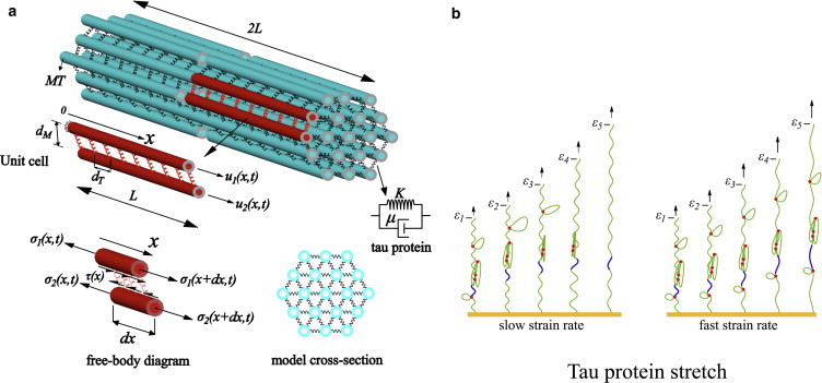
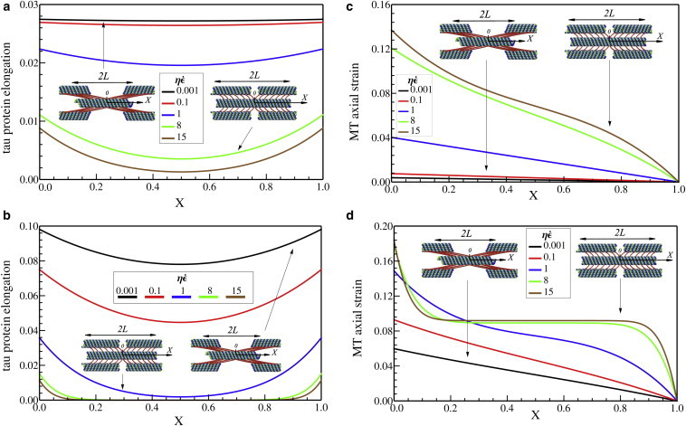
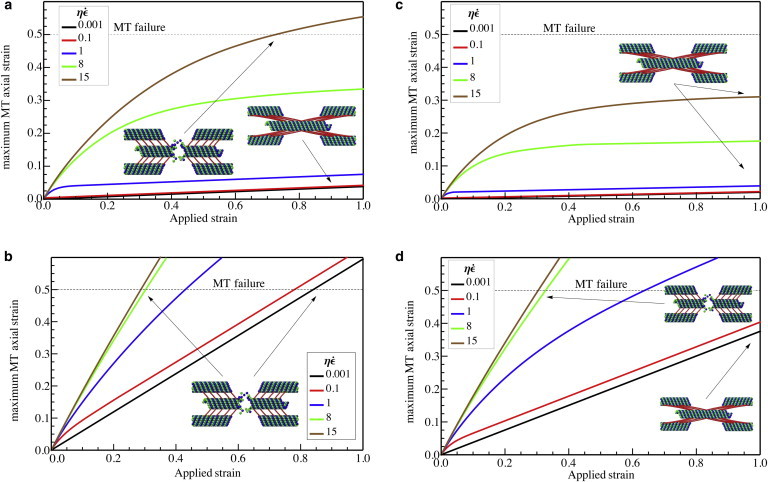
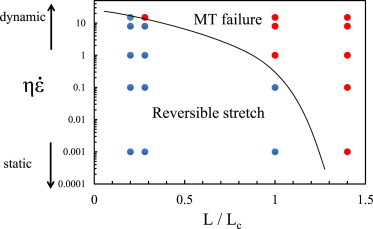
The unique viscoelastic nature of axons is thought to underlie selective vulnerability to damage during traumatic brain injury. In particular, dynamic loading of axons has been shown to mechanically break microtubules at the time of injury. However, the mechanism of this rate-dependent response has remained elusive. Here, we present a microstructural model of the axonal cytoskeleton to quantitatively elucidate the interaction between microtubules and tau proteins under mechanical loading. Mirroring the axon ultrastructure, the microtubules were arranged in staggered arrays, cross-linked by tau proteins. We found that the viscoelastic behavior specifically of tau proteins leads to mechanical breaking of microtubules at high strain rates, whereas extension of tau allows for reversible sliding of microtubules without any damage at small strain rates. Based on the stiffness and viscosity of tau proteins inferred from single-molecule force spectroscopy studies, we predict the critical strain rate for microtubule breaking to be in the range 22-44 s(-1), in excellent agreement with recent experiments on dynamic loading of micropatterned neuronal cultures. We also identified a characteristic length scale for load transfer that depends on microstructural properties and have derived a phase diagram in the parameter space spanned by loading rate and microtubule length that demarcates those regions where axons can be loaded and unloaded reversibly and those where axons are injured due to breaking of the microtubules.


Figures

Figure 1
Figure 1
(A) Electron microscopy image of porcine brain MTs polymerized with tau protein. (Arrows) Cross-linkers between the MTs. Scale bar = 100 nm. (Reprinted with permission from ©1988 Rockefeller University Press. Originally published in Hirokawa et al. (8).) (B) Experimental results for injury of cultured 2-mm-long axons (5). A controlled air pulse (with duration <50 ms) is applied to the axon and the increase in the length is used to infer tensile stretch of the axon. (Arrows) Intact straight MTs inside the axon; (asterisk) broken ends of the MTs at the peak of the generated undulations. (C) Due to the interruption of the axonal transport, swelling starts to appear at the end of the broken MTs (asterisks) (D) The swelling grows and becomes more apparent in the transmission electron microscopy micrographs (taken at 1–2 h after injury). Scale bars in panels BD = 500 nm. Reprinted with permission from Tang-Schomer et al. (5).
Figure 2
Figure 2
(a) The microstructure of the axon consists of MTs cross-linked by tau proteins (springs). The unit cell used in the mathematical analysis with staggered neighbors is also shown. The viscoelastic model considered for the tau protein consists of a spring (stiffness K) and a dashpot (viscosity μ). (Free-body diagram) Axial and shear components of forces acting on the MTs. (b) The response of the tau protein to different pulling rates at five different strains, ε1ε5. (Red dots) Intramolecular bonds; (green solid-curve) backbone of the tau protein. At slow strain rates, the intramolecular bonds rupture at smaller force levels (magnitude of the force is proportional to arrow lengths), whereas at fast strain rates, larger force levels are needed to stretch the protein. (Blue segment) Section of the protein backbone that stretches at fast rates of pulling, whereas its length is nearly constant at slow rates. To see this figure in color, go online.
Figure 3
Figure 3
Elongation of tau proteins along the MTs, U2(X,T) – U1(X,T), with (a) L/Lc = 0.28 and (b) L/Lc = 1.40 and also axial strain along microtubules, ∂U1(X,T)/∂X, with (c) L/Lc = 0.28 and (d) L/Lc = 1.40, when overall elongation of the axon is 10%. The centers of the MTs stretch more with increasing strain rate and increasing length. To see this figure in color, go online.
Figure 4
Figure 4
The maximum axial strain in the MTs, when the spacing between the tau proteins is 30 nm, with (a) L = 1 μm, L/Lc = 0.28 and (b) L = 5 μm, L/Lc = 1.40 and also when the spacing between the tau proteins is increased to 60 nm with (c) L = 1 μm, L/Lc = 0.20 and (d) L = 5 μm, L/Lc = 1.00, as a function of the total axonal strain. To see this figure in color, go online.
Figure 5
Figure 5
Phase-diagram that demarcates regions where MTs break and where axon can be reversibly deformed. The symbols (red = MT breaking, blue = MT sliding) are from numerical solutions presented in Fig. 4. To see this figure in color, go online.

References

    1. Johnson V.E., Stewart W., Smith D.H. Axonal pathology in traumatic brain injury. Exp. Neurol. 2013;246:35–43. - PMC - PubMed
    1. Knöferle J., Koch J.C., Lingor P. Mechanisms of acute axonal degeneration in the optic nerve in vivo. Proc. Natl. Acad. Sci. USA. 2010;107:6064–6069. - PMC - PubMed
    1. Cloots R.J.H., van Dommelen J.A.W., Geers M.G.D. Multi-scale mechanics of traumatic brain injury: predicting axonal strains from head loads. Biomech. Model. Mechanobiol. 2013;12:137–150. - PubMed
    1. Smith D.H., Meaney D.F. Axonal damage in traumatic brain injury. Neuroscience. 2000;6:483–495.
    1. Tang-Schomer M.D., Patel A.R., Smith D.H. Mechanical breaking of microtubules in axons during dynamic stretch injury underlies delayed elasticity, microtubule disassembly, and axon degeneration. FASEB J. 2010;24:1401–1410. - PMC - PubMed

Publication types