Delayed formation of zero-valent selenium nanoparticles by Bacillus mycoides SeITE01 as a consequence of selenite reduction under aerobic conditions
- PMID: 24606965
- PMCID: PMC3975340
- DOI: 10.1186/1475-2859-13-35
Delayed formation of zero-valent selenium nanoparticles by Bacillus mycoides SeITE01 as a consequence of selenite reduction under aerobic conditions
Abstract
Background: Selenite (SeO32-) oxyanion shows severe toxicity to biota. Different bacterial strains exist that are capable of reducing SeO32- to non-toxic elemental selenium (Se0), with the formation of Se nanoparticles (SeNPs). These SeNPs might be exploited for technological applications due to their physico-chemical and biological characteristics. The present paper discusses the reduction of selenite to SeNPs by a strain of Bacillus sp., SeITE01, isolated from the rhizosphere of the Se-hyperaccumulator legume Astragalus bisulcatus.
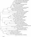
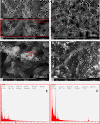
Results: Use of 16S rRNA and GyrB gene sequence analysis positioned SeITE01 phylogenetically close to B. mycoides. On agarized medium, this strain showed rhizoid growth whilst, in liquid cultures, it was capable of reducing 0.5 and 2.0 mM SeO32- within 12 and 24 hours, respectively. The resultant Se0 aggregated to form nanoparticles and the amount of Se0 measured was equivalent to the amount of selenium originally added as selenite to the growth medium. A delay of more than 24 hours was observed between the depletion of SeO32 and the detection of SeNPs. Nearly spherical-shaped SeNPs were mostly found in the extracellular environment whilst rarely in the cytoplasmic compartment. Size of SeNPs ranged from 50 to 400 nm in diameter, with dimensions greatly influenced by the incubation times. Different SeITE01 protein fractions were assayed for SeO32- reductase capability, revealing that enzymatic activity was mainly associated with the membrane fraction. Reduction of SeO32- was also detected in the supernatant of bacterial cultures upon NADH addition.
Conclusions: The selenite reducing bacterial strain SeITE01 was attributed to the species Bacillus mycoides on the basis of phenotypic and molecular traits. Under aerobic conditions, the formation of SeNPs were observed both extracellularly or intracellularly. Possible mechanisms of Se0 precipitation and SeNPs assembly are suggested. SeO32- is proposed to be enzymatically reduced to Se0 through redox reactions by proteins released from bacterial cells. Sulfhydryl groups on peptides excreted outside the cells may also react directly with selenite. Furthermore, membrane reductases and the intracellular synthesis of low molecular weight thiols such as bacillithiols may also play a role in SeO32- reduction. Formation of SeNPs seems to be the result of an Ostwald ripening mechanism.
Figures


Similar articles
-
Untargeted Metabolomics Investigation on Selenite Reduction to Elemental Selenium by Bacillus mycoides SeITE01.Front Microbiol. 2021 Sep 16;12:711000. doi: 10.3389/fmicb.2021.711000. eCollection 2021. Front Microbiol. 2021. PMID: 34603239 Free PMC article.
-
Reduction of selenite to Se(0) nanoparticles by filamentous bacterium Streptomyces sp. ES2-5 isolated from a selenium mining soil.Microb Cell Fact. 2016 Sep 15;15(1):157. doi: 10.1186/s12934-016-0554-z. Microb Cell Fact. 2016. PMID: 27630128 Free PMC article.
-
Selenite biotransformation and detoxification by Stenotrophomonas maltophilia SeITE02: Novel clues on the route to bacterial biogenesis of selenium nanoparticles.J Hazard Mater. 2017 Feb 15;324(Pt A):3-14. doi: 10.1016/j.jhazmat.2016.02.035. Epub 2016 Feb 16. J Hazard Mater. 2017. PMID: 26952084
-
Biogenic selenium nanoparticles: current status and future prospects.Appl Microbiol Biotechnol. 2016 Mar;100(6):2555-66. doi: 10.1007/s00253-016-7300-7. Epub 2016 Jan 22. Appl Microbiol Biotechnol. 2016. PMID: 26801915 Review.
-
Effects of selenium nanoparticles produced by Lactobacillus acidophilus HN23 on lipid deposition in WRL68 cells.Bioorg Chem. 2024 Apr;145:107165. doi: 10.1016/j.bioorg.2024.107165. Epub 2024 Feb 7. Bioorg Chem. 2024. PMID: 38367427 Review.
Cited by
-
Selenium nanoparticles alleviate renal ischemia/reperfusion injury by inhibiting ferritinophagy via the XBP1/NCOA4 pathway.Cell Commun Signal. 2024 Jul 25;22(1):376. doi: 10.1186/s12964-024-01751-2. Cell Commun Signal. 2024. PMID: 39061070 Free PMC article.
-
A Comparative Study of the Synthesis and Characterization of Biogenic Selenium Nanoparticles by Two Contrasting Endophytic Selenobacteria.Microorganisms. 2023 Jun 16;11(6):1600. doi: 10.3390/microorganisms11061600. Microorganisms. 2023. PMID: 37375102 Free PMC article.
-
Characterization and biological activity of selenium nanoparticles biosynthesized by Yarrowia lipolytica.Microb Biotechnol. 2024 Oct;17(10):e70013. doi: 10.1111/1751-7915.70013. Microb Biotechnol. 2024. PMID: 39364622 Free PMC article.
-
Unveiling the innovative green synthesis mechanism of selenium nanoparticles by exploiting intracellular protein elongation factor Tu from Bacillus paramycoides.J Zhejiang Univ Sci B. 2024 Aug 19;25(9):789-795. doi: 10.1631/jzus.B2300738. J Zhejiang Univ Sci B. 2024. PMID: 39308068 Free PMC article.
-
High-Efficiency Reducing Strain for Producing Selenium Nanoparticles Isolated from Marine Sediment.Int J Mol Sci. 2022 Oct 8;23(19):11953. doi: 10.3390/ijms231911953. Int J Mol Sci. 2022. PMID: 36233255 Free PMC article.
References
-
- Fordyce FM. Selenium deficiency and toxicity in the environment. Essent Med Geology. 2013;16:375–416.
-
- Ralston NVC, Ralston CR, Blackwell JL III, Raymond LJ. Dietary and tissue selenium in relation to methylmercury toxicity. Neurotoxicology. 2008;29:802.811. - PubMed
-
- Keller EA. Environmental Geology. 9. Upper Saddle River, NJ, USA: Prentice Hall; 2000.
-
- Craig PJ, Maher W. In: Organometallic Compounds in the Environment. 2. Craig PJ, editor. Chichester: Wiley; 2003. Organoselenium compounds in the environment; pp. 391–398.
Publication types
MeSH terms
Substances
LinkOut - more resources
Full Text Sources
Other Literature Sources

