Review
. 2014 Mar 5;81(5):967-983.
doi: 10.1016/j.neuron.2014.02.025.

Optic ataxia: from Balint's syndrome to the parietal reach region

Affiliations
Review

Optic ataxia: from Balint's syndrome to the parietal reach region

Richard A Andersen et al. Neuron. .

Abstract

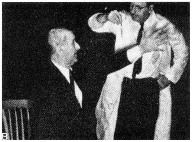
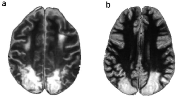
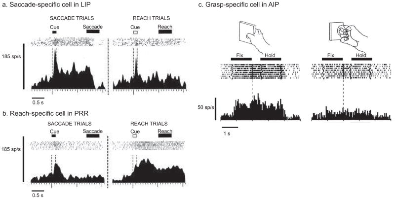
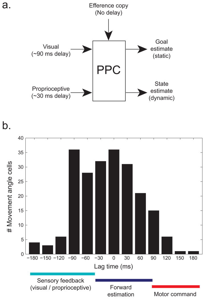
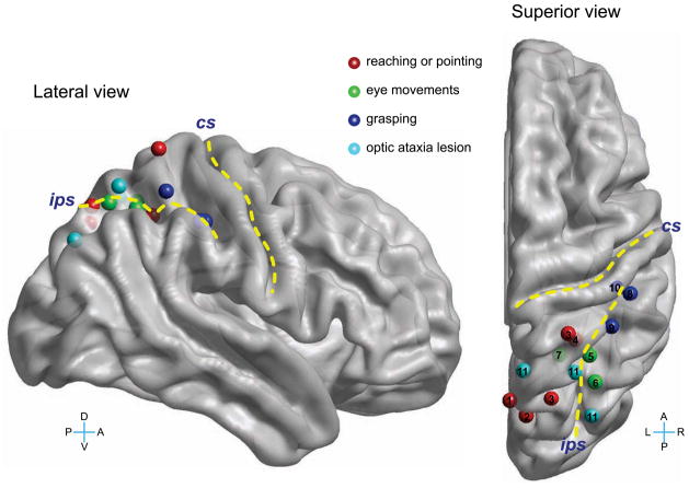
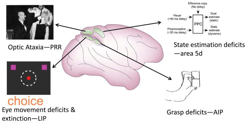
Optic ataxia is a high-order deficit in reaching to visual goals that occurs with posterior parietal cortex (PPC) lesions. It is a component of Balint's syndrome that also includes attentional and gaze disorders. Aspects of optic ataxia are misreaching in the contralesional visual field, difficulty preshaping the hand for grasping, and an inability to correct reaches online. Recent research in nonhuman primates (NHPs) suggests that many aspects of Balint's syndrome and optic ataxia are a result of damage to specific functional modules for reaching, saccades, grasp, attention, and state estimation. The deficits from large lesions in humans are probably composite effects from damage to combinations of these functional modules. Interactions between these modules, either within posterior parietal cortex or downstream within frontal cortex, may account for more complex behaviors such as hand-eye coordination and reach-to-grasp.


Figures

Figure 1
Figure 1. Patient with optic ataxia
The patient misreaches beyond the pencil when asked to touch it. From Cogan 1965.
Figure 2
Figure 2. Patient lesions are large and likely involve several functional areas
Horizontal magnetic resonance imagining sections through the bilateral parietal lobe lesions of patients A.T. (a) and I.G. (b). From Schindler et al. 2004.
Figure 3
Figure 3. Action specificity in non-human primate posterior parietal areas
a–b. Cells recorded from the lateral intraparietal area (LIP) and the parietal reach region (PRR) in saccade versus reach trials. The LIP neuron is more active in saccade trials, whereas the PRR neuron is more active in reach trials. Modified from Snyder et al. 1997. c. Cell recorded from the anterior intraparietal area (AIP) for different objects and hand manipulation. This visuomotor neuron is highly selective for both object and grip type. Modified from Murata et al. 2000.
Figure 4
Figure 4. State estimation in area 5
a. Model of posterior parietal cortex sensorimotor integration. Sensory signals arrive with a delay, approximately 90 ms for visual inputs and 30ms for proprioceptive inputs. An efference copy signal of movement commands arrives with no delay. Outputs include a movement-goal location estimate and a dynamic estimate of the current movement state. b. Histogram of optimal lag times for area 5 cells sensitive to the movement angle. The lag times are the times at which the cells best estimate the instantaneous movement angle. Overall cells show a distribution of lag times that are centered around 0 ms, consistent with the forward state estimation hypothesis. Modified from Mulliken et al. 2008.
Figure 5
Figure 5. Action specificity in human posterior parietal areas
Lesion sites in optic ataxia and action specific sites for reaching or pointing, eye movements, and grasping. The yellow lines demarcate the central (CS) and intraparietal sulci (IPS). L, R, D, V are left, right, dorsal, ventral. The Talairach coordinates of these sites, reported in Culham et al. 2006, were translated to the Montreal Neurological Institute template, and then visualized using BrainNet Viewer (Xia et al. 2013). Not all sites are clearly visible in the lateral view due to the convolutions of the brain. The numbers in the superior view indicate the original studies that identified these sites: 1) Connolly et al. 2003, 2) Astafiev et al. 2003, 3) Prado, et al. 2005, 4) Grefkes et al. 2004, 5) Petit et al. 1999, 6) Sereno et al. 2001, 7) Medendorp et al. 2003, 8) Binkofski et al. 1998, 9) Culham et al. 2003, 10) Frey et al. 2005, 11) Karnath & Perenin 2005.
Figure 6
Figure 6. Optic ataxia after parietal reach region inactivation
a. Coronal MRI slice through the injection site in PRR in monkey Y. The injection site appears bright due to injection of the contrast agent gadolinium. The dotted blue line indicates the intraparietal sulcus. b. Movement amplitudes for reaches and saccades in control (black) and inactivation (purple) sessions. The reach amplitudes are hypometric but saccades are unaffected. c. Reaches to extrafoveal targets are affected but not reaches to foveal targets. Modified from Hwang et al. 2012.
Figure 7
Figure 7. Extinction after lateral intraparietal area inactivation
a. Coronal MRI slice through the injection site in LIP of monkey F. b. Schematic of the choice task. The animal is free to choose between targets in the ipsilesional and contralesional visual fields. The red square is the fixation point and purple squares the two saccade targets. c. Percent of choices into the ipsilesional field. In the control condition (black) the animal chooses nearly equally between fields but in the inactivation condition (purple) the animal is biased toward the ipsilesional target. Modified from Wilke et al. 2012.
Figure 8
Figure 8. Proposed functional segregation within posterior parietal cortex
Area PRR inactivation produces optic ataxia, LIP inactivation eye movement deficits and extinction, area 5d inactivation online deficits consistent with disruption of state estimation, and AIP grasp deficits. These inactivation results and recording data suggest specialization of PRR for reach, LIP for eye movements and attention, area 5d for state estimation, and AIP for grasp.

References

    1. Amin H, Ehrreich S, Kelly J, Lebby P, Hutchinson H. Balint syndrome in the pediatric population: A case report of three patients. J Neurol. 2012:P02.179.
    1. Andersen RA, Essick GK, Siegel RM. The encoding of spatial location by posterior parietal neurons. Science. 1985;230:456–458. - PubMed
    1. Andersen RA, Essick GK, Siegel RM. Neurons of area 7 activated by both visual stimuli and oculomotor behavior. Exp Brain Res. 1987;67:316–322. - PubMed
    1. Andersen RA, Bracewell RM, Barash S, Gnadt JW, Fogassi L. Eye position effects on visual, memory and saccade-related activity in areas LIP and 7A of macaque. J Neurosci. 1990;10:1176–1196. - PMC - PubMed
    1. Andersen RA, Buneo CA. Intentional maps in posterior parietal cortex. Ann Rev Neurosci. 2002;25:189–220. - PubMed

Publication types

LinkOut - more resources