. 2014 Mar 6;94(3):470-8.
doi: 10.1016/j.ajhg.2014.02.004.

Mutations in USP9X are associated with X-linked intellectual disability and disrupt neuronal cell migration and growth

Affiliations

Mutations in USP9X are associated with X-linked intellectual disability and disrupt neuronal cell migration and growth

Claire C Homan et al. Am J Hum Genet. .

Abstract

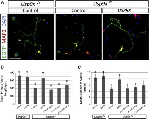
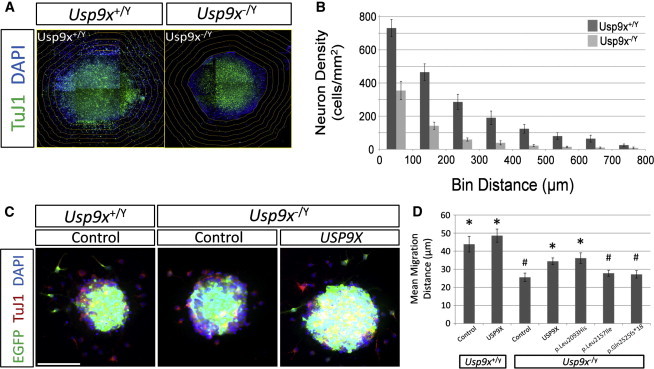
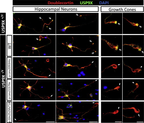
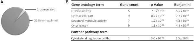
With a wealth of disease-associated DNA variants being recently reported, the challenges of providing their functional characterization are mounting. Previously, as part of a large systematic resequencing of the X chromosome in 208 unrelated families with nonsyndromic X-linked intellectual disability, we identified three unique variants (two missense and one protein truncating) in USP9X. To assess the functional significance of these variants, we took advantage of the Usp9x knockout mouse we generated. Loss of Usp9x causes reduction in both axonal growth and neuronal cell migration. Although overexpression of wild-type human USP9X rescued these defects, all three USP9X variants failed to rescue axonal growth, caused reduced USP9X protein localization in axonal growth cones, and (in 2/3 variants) failed to rescue neuronal cell migration. Interestingly, in one of these families, the proband was subsequently identified to have a microdeletion encompassing ARID1B, a known ID gene. Given our findings it is plausible that loss of function of both genes contributes to the individual's phenotype. This case highlights the complexity of the interpretations of genetic findings from genome-wide investigations. We also performed proteomics analysis of neurons from both the wild-type and Usp9x knockout embryos and identified disruption of the cytoskeleton as the main underlying consequence of the loss of Usp9x. Detailed clinical assessment of all three families with USP9X variants identified hypotonia and behavioral and morphological defects as common features in addition to ID. Together our data support involvement of all three USP9X variants in ID in these families and provide likely cellular and molecular mechanisms involved.


Figures

Figure 1
Figure 1
Expression of USP9X but Not USP9X Variants Rescues Axonal Defects in Hippocampal Neurons Isolated from Usp9x Knockout Mice Usp9xloxP/LoxP female mice were crossed with Nestin-Cre males to delete Usp9x from the entire brain from E12.5 onward as previously described. Brains of male embryos from these matings are either wild-type (Usp9x+/Y) or knockout (Usp9x−/Y). For all experiments, hippocampal neurons were isolated from male embryos at E18.5 by established methods and nucleofected with 1 μg of pMAX-EGFP in addition to either 5 μg of the empty expression vector (pCMV; Control) or expression vectors containing USP9X (pCMV-USP9X (USP9X)) or USP9X variants (pCMV-USP9X_Leu2093His, pCMV-USP9X_Leu2157Ile, or pCMV-USP9X_Gln2525fs18) as previously described. After nucleofection, neurons were seeded in vitro and grown for 5 days, fixed and immunofluorescently stained, and subjected to morphometric analysis as previously described. (A) Representative immunofluorescent images of wild-type neurons nucleofected with control plasmids, compared with knockout neurons nucleofected with either control plasmid or USP9X expression plasmid. Nucleofected neurons are identified by EGFP expression (green). Scale bar represents 100 μm. (B) Loss of Usp9x reduces axonal length, which is rescued by overexpression of USP9X but not USP9X variants. Morphometric analysis of mean axon length conducted as previously described. Note that overexpression of USP9X in wild-type neurons has no significant effect. (C) Loss of Usp9x reduces the number of axonal termini, which is rescued by overexpression of USP9X but not USP9X variants. Morphometric analysis of mean axonal termini was conducted as previously described. Note that overexpression of USP9X in wild-type neurons has no significant effect. All experiments were conducted in triplicate, with at least 25 neurons scored per replicate (i.e., >75 neurons scored per experimental condition). Graphed values are the mean of the three average values derived from each replicate. Error bars are ±SD. Statistical analysis conducted by Student’s two-tailed, unpaired t test, assuming equal variance, and significance set as p < 0.05: significantly different to “Usp9x−/Y + Control” condition; #significantly different to “Usp9x−/Y+ USP9X” condition.
Figure 2
Figure 2
Expression of USP9X but Not USP9X Variants Rescues Neuronal Migration Defects in Cells Isolated from Usp9x Knockout Mice Wild-type (Usp9x+/Y) or knockout (Usp9x−/Y) embryonic brains were generated and isolated as described earlier (see Figure 1). Neural progenitor cells (NPCs) were isolated from the dorsal cortex of E18.5 brains and grown as nonadherent neurospheres in culture as previously described. (A and B) Neuronal migration away from seeded neurospheres is inhibited in the absence of Usp9x. Passage 3 neurospheres were cultured for 5 days, adhered to a poly-L-lysine surface substrate (seeding) as previously described, cultured a further 5 days, fixed, and immunofluorescently stained as previously described. (A) Scoring system used to measure neuronal cell migration from seeded neurospheres. Composite images of immunofluorescently stained neurons present in neurosphere outgrowths (TuJ1: anti-βIII tubulin, green; DAPI: blue) were collated and the neurosphere periphery (as indicated by the edge of contiguous nuclei) outlined. The outline was then used to generate concentric bins outside of the neurosphere (i.e., in the migration zones) of increasing size (increases of 100 μm diameter) via ImageJ (NIH) software tools. This allowed for the calculation of neuronal cell density in each bin, which thus reports on migration independent of initial neurosphere size, and of bin volumes. (B) Quantitation of neuronal migration away from seeded neurospheres by the described scoring system. Neurospheres were isolated from three wild-type and three Usp9x knockout embryos. Scoring was restricted to spheres of similar size. At least five neurosphere outgrowths were scored per embryo (total outgrowths scored: wild-type, 32; knockout, 16). Graph values are means and error bars are ±SD, of pooled data. (C and D) Expression of USP9X but not USP9X variants rescue neuronal migration defects. Neurospheres were nucleofected via the same regime described in Figure 1, with previously described nucleofection techniques. Neurospheres were allowed to recover for 48 hr prior to seeding and were cultured for an additional 48 hr to promote neuronal migration, prior to fixing for immunofluorescent staining as described above. (C) Representative images showing migration from wild-type neurospheres nucleofected with control plasmids, compared with Usp9x knockout neurospheres nucleofected with either control plasmid or USP9X expression plasmid. Scale bar represents 100 μm. (D) To measure the migration of nucleofected neurons after 2 days of adherent culture, an alternative scoring system was adopted. Individually nucleofected neurons (i.e., expressing EGFP [green] and labeled by TuJ1 antibody [red]) were scored for the length of their radial migration path extending from the periphery of their sphere of origin. The average migration distance of neurons was calculated per neurosphere, and the mean migration distance of all neurospheres per condition was calculated and graphed. At least 20 neurospheres were scored per condition. Error bars represent ±SD. Statistical analysis conducted by Student’s two-tailed, unpaired t test, assuming equal variance, and significance set as p < 0.05: significantly different to “Usp9x−/Y + Control” condition; #significantly different to “Usp9x/Y+ USP9X” condition.
Figure 3
Figure 3
Reduced Localization of Variant USP9X in Axonal Growth Cones Wild-type (Usp9x+/Y) or knockout (Usp9x−/Y) hippocampal neuronal cultures were generated and nucleofected with USP9X or variant forms as described previously (Figure 1). Immunoflourescent staining of Doublecortin (Dcx; red) and USP9X (green). Cell nuclei were stained with DAPI (blue). Open arrows indicate USP9X enrichment and closed arrows indicate depletion of USP9X in axonal growth cones. Scale bars represent 50 μm (left panels) and 20 μm (right panels).
Figure 4
Figure 4
Loss of Usp9x Disrupts Cytoskeleton Components Cortical neurons were isolated from either wild-type (Usp9x+/Y; n = 4) or knockout (Usp9x−/Y; n = 4) E18.5 embryos and cultured in vitro as previously described. After 5 days, cell lysates were generated and differentially expressed proteins identified as described in the text. (A) Loss of Usp9x downregulates proteins. A total of 28 unique proteins were identified as being deregulated, with 27 of them downregulated in the absence of Usp9x. (B) Gene ontology and PANTHER pathway analysis summary. The list of deregulated genes was analyzed by DAVID.

References

    1. Ropers H.H. Genetics of early onset cognitive impairment. Annu. Rev. Genomics Hum. Genet. 2010;11:161–187. - PubMed
    1. Gécz J., Shoubridge C., Corbett M. The genetic landscape of intellectual disability arising from chromosome X. Trends Genet. 2009;25:308–316. - PubMed
    1. Tarpey P.S., Smith R., Pleasance E., Whibley A., Edkins S., Hardy C., O’Meara S., Latimer C., Dicks E., Menzies A. A systematic, large-scale resequencing screen of X-chromosome coding exons in mental retardation. Nat. Genet. 2009;41:535–543. - PMC - PubMed
    1. Whibley A.C., Plagnol V., Tarpey P.S., Abidi F., Fullston T., Choma M.K., Boucher C.A., Shepherd L., Willatt L., Parkin G. Fine-scale survey of X chromosome copy number variants and indels underlying intellectual disability. Am. J. Hum. Genet. 2010;87:173–188. - PMC - PubMed
    1. Shoubridge C., Tarpey P.S., Abidi F., Ramsden S.L., Rujirabanjerd S., Murphy J.A., Boyle J., Shaw M., Gardner A., Proos A. Mutations in the guanine nucleotide exchange factor gene IQSEC2 cause nonsyndromic intellectual disability. Nat. Genet. 2010;42:486–488. - PMC - PubMed

Publication types

MeSH terms