Review
. 2014 Mar 6;14(3):306-21.
doi: 10.1016/j.stem.2014.02.002.

Metastatic stem cells: sources, niches, and vital pathways

Affiliations
Review

Metastatic stem cells: sources, niches, and vital pathways

Thordur Oskarsson et al. Cell Stem Cell. .

Abstract

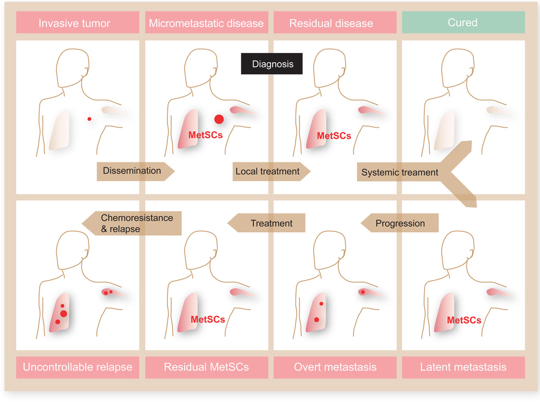
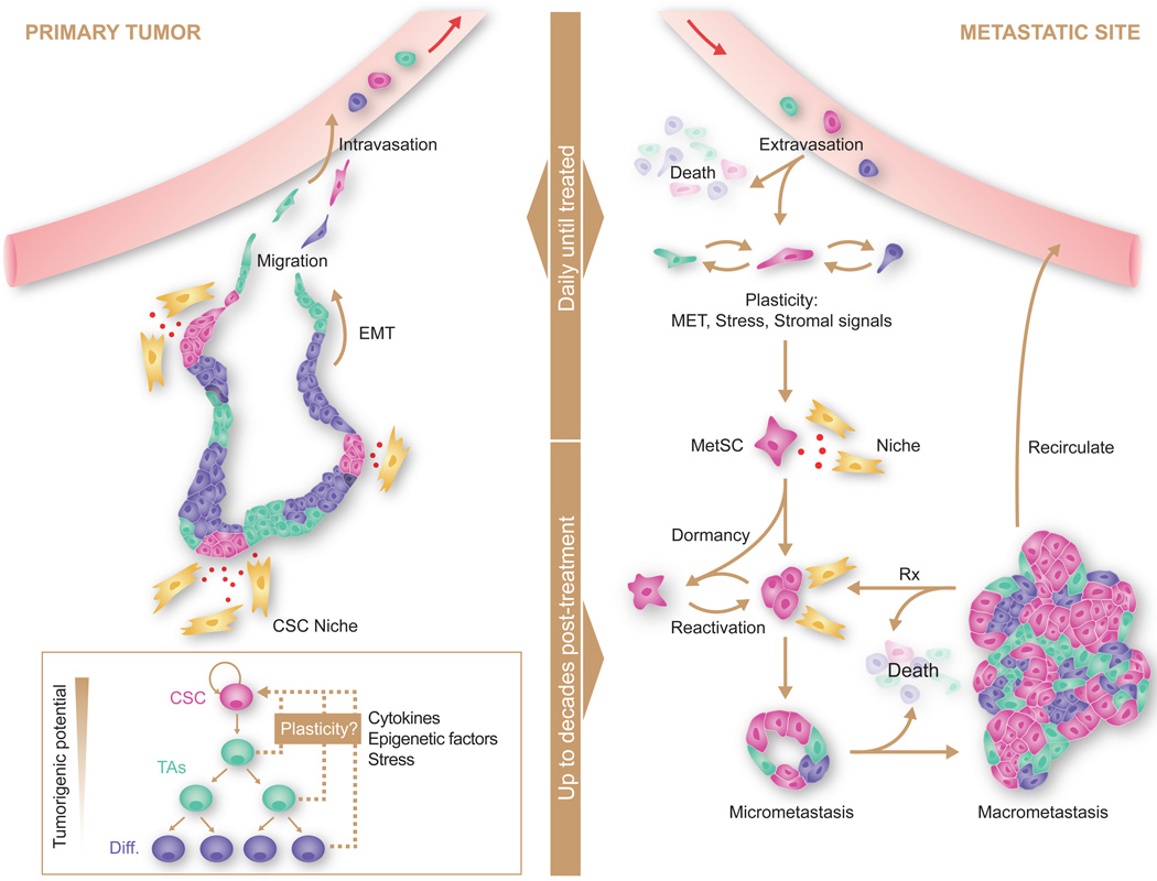
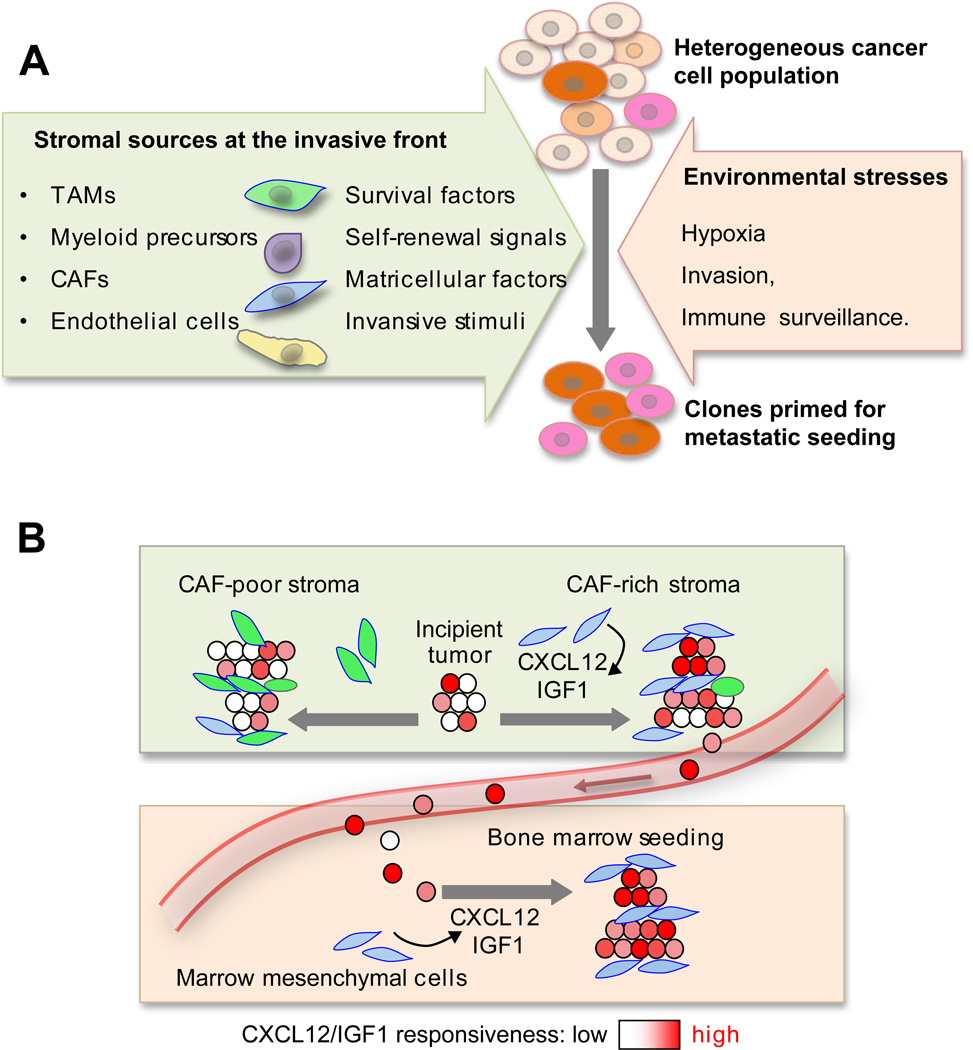
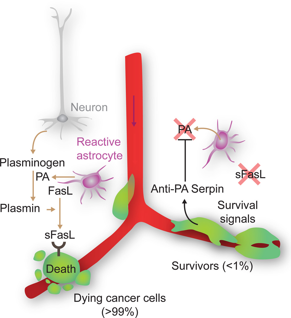
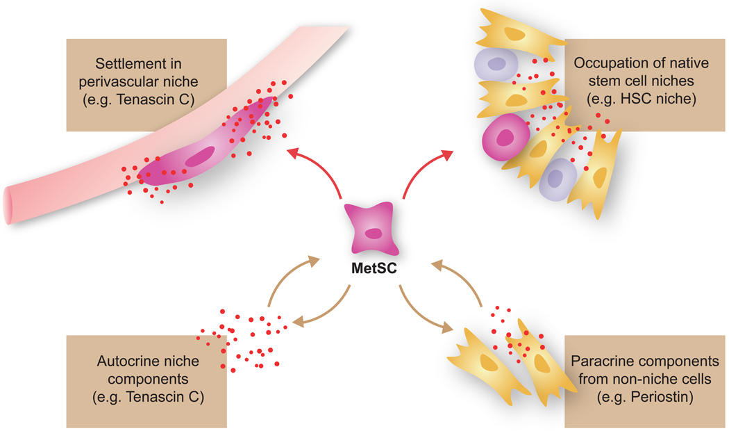
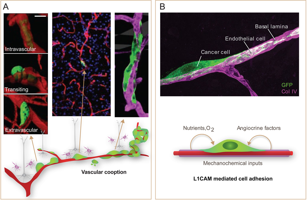
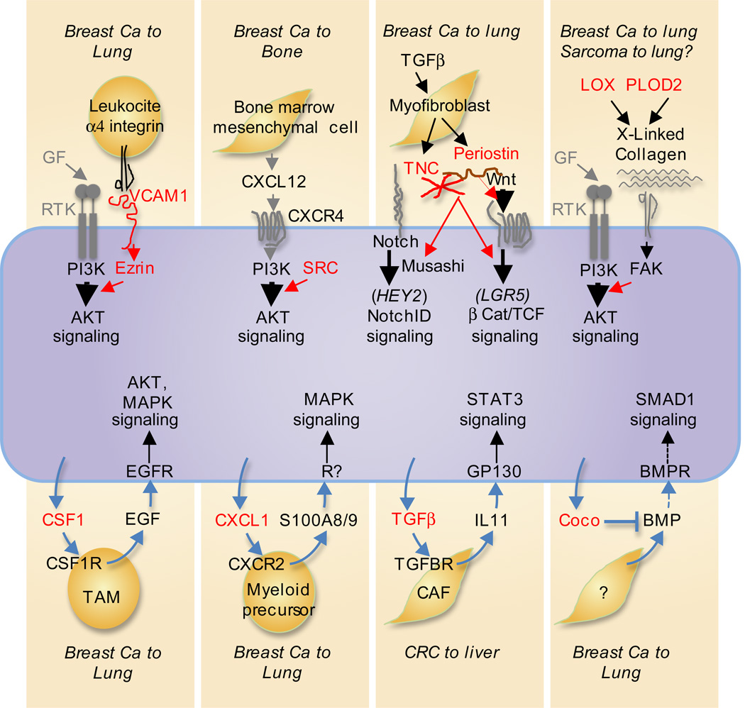
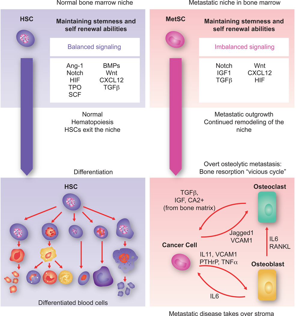
Metastasis is powered by disseminated cancer cells that re-create a full-fledged tumor in unwelcoming tissues, away from the primary site. How cancer cells moving from a tumor into the circulation manage to infiltrate distant organs and initiate metastatic growth is of interest to cancer biologists and clinical oncologists alike. Recent findings have started to define the sources, phenotypic properties, hosting niches, and signaling pathways that support the survival, self-renewal, dormancy, and reactivation of cancer cells that initiate metastasis: metastatic stem cells. By dissecting the biology of this process, vulnerabilities are being exposed that could be exploited to prevent metastasis.


Figures

Figure 1
Figure 1. Deadly seeds left behind in a typical course of metastatic cancer
At diagnosis a primary tumor (a carcinoma of the lung or breast in this example) may have already seeded distant organs with cancer cells, including cells with tumor-initiating capacity that are defined here as metastatic stem cells (MetSCs). After diagnosis, the primary tumor may be removed by surgery and irradiation, and disseminated cancer cells may be eliminated by systemic chemotherapy, leading to a cure. Alternatively, residual MetSCs may remain in a latent state, eventually giving rise to overt metastasis. New rounds of therapy may then induce regression of the metastatic lesions, but chemoresistant MetSCs selected during each round of treatment may eventually give rise to uncontrollable metastasis. This process is responsible for 90% of deaths from cancer.
Figure 2
Figure 2. Sources, dissemination, dormancy, and outgrowth of MetSCs
Several types of cancer display a hierarchical organization with CSCs being the only cell type with long-term self-renewal potential. The progeny of CSCs –the transit amplifying progenitors (TAs) and their differentiated derivatives– are short lived and have a lower tumorigenic potential. Conversion of non-CSCs into CSCs occurs in certain types of cancer and can be triggered by cytokines and stress conditions. CSCs interact with microenvironmental niches that sustain the tumor-perpetuating potential of the cells. Both CSCs and non-CSCs can display migratory behavior at the invasive front of primary tumors, frequently associated with an EMT. Intravasation, circulation, and extravasation of cancer cells in these various states can occur continuously until the primary tumor is removed. Survival of disseminated cancer cells at distant sites is a limiting step, as the vast majority of infiltrating cancer cells die. The survivors that are endowed with tumor-initiating capacity constitute MetSCs. Cells that had previously undergone an EMT must reacquire epithelial traits and co-opt a supportive stromal niche in order to thrive in the new environment. Additionally, disseminated non-CSCs may convert into MetSCs through still poorly understood processes of phenotypic plasticity. MetSCs may generate progeny and give rise to overt metastasis right after infiltrating the host tissue. More frequently, however, disseminated cancer cells enter a dormant state that can last for decades and is largely resistant to current therapies. Upon exit from quiescence, MetSCs may regenerate their lineage in the host organ and release metastatic progeny into the circulation to start secondary lesions in the same or other organs. Treatment of overt metastasis seldom results in eradication of the disease, as residual MetSCs frequently regenerate the tumor after each drug treatment cycle.
Figure 3
Figure 3. Selection of metastatic traits in primary tumors
A. Cancer cells at the invasive front of primary tumors are exposed to the stresses of invading surrounding tissue, hypoxia, and immune surveillance. Various stromal cell types produce signals that cancer cells can use for survival, self-renewal, invasiveness and migration. Under selective pressure, these signals skew the heterogeneous cancer cell population towards a preponderance of clones that are primed for survival also under stress of infiltrating distant tissues. B. When the stroma of a primary tumor is rich in cell and signals that resemble those of a particular distant tissue, cancer cell clones selected in the primary tumor may be primed to thrive in that particular tissue. An example is provided by the ability of a CAF-rich stroma in breast tumors to select for cancer cell clones that are fittest to respond to the CAF-derived factors CXCL12 and IGF1, and thereby primed for survival in the CXCL12- and IGF1-rich environment of the bone marrow. [Image adapted from (Zhang et al., 2013b)].
Figure 4
Figure 4. A reactive stroma kills infiltrating cancer cells
Astrocytes in the brain stroma react to infiltrating breast or lung cancer cells by expressing plasminogen activator (PA). PA generates plasmin that cleaves and mobilizes membrane-bound Fas ligand (FasL) from astrocytes. The cancer cells succumb to Fas-mediated apoptosis, but can avert this fate by expressing PA inhibitory serpins. Neuroserpin is normally expressed only in neurons, which use it for protection against astrocytes reacting to brain injury. [Image adapted from (Valiente et al., 2014)].
Figure 5
Figure 5. Three possible sources of metastatic niche support
Disseminated cancer cells can obtain stem cell niche support by occupying native stem cell niches including perivascular sites, by recruiting stromal cells that produce stem cell niche-like components, or by producing niche components themselves.
Figure 6
Figure 6. The perivascular niche for metastasis initiation
A. Metastasis-initiating cells (green) exiting from brain capillaries (red) remain tightly associated with the vessels, adhering to and stretching over their abluminal surface. This interaction is required for metastatic outgrowth. Outgrowth occurs forming a furrow over the capillary and later a multilayered cell colony. B. The axonal guidance cell adhesion receptor L1CAM is aberrantly expressed in tumors, and its expression is associated with relapse. L1CAM in metastasis-initiating cells (green) that infiltrate the brain mediates their adhesion to capillary basal lamina (magenta). Besides providing MetSCs with mechanochemical cues, vascular cooption facilitates their access to oxygen and nutrients from the blood supply and to factors from the endothelium and surrounding stroma. [Adapted from (Valiente et al., 2014)].
Figure 7
Figure 7. Pathway amplifiers and paracrine loops for MetSC support
Many of the known traits that promote metastatic seeding involve gene products (red) that the cancer cells express to amplify their own responsiveness to vital stromal cues. The cues that activate these pathways include receptor tyrosine kinase (RTK) growth factor ligands (GF), chemokines like CXCL12, and Wnt, and Notch ligands. Examples amplifiers include cell adhesion receptors like the VCAM1-Ezrin complex that is engaged by tumor leukocyte integrins; the tyrosine kinase Src that amplifies PI3K-AKT activation by CXCR4 and IGF1R (not shown); the cancer cell- and myofibroblast-derived ECM components tenascin C (TNC) and periostin that enhance Wnt access to their receptors (periostin) or amplify signaling by the Wnt and Notch pathways (TNC); and, the collagen crosslinking enzymes LOX and PLOD2 that stiffen the ECM for integrin/focal adhesion kinase (FAK) mediated amplification of RTK signaling. Various cancer cell-derived cytokines (bottom, red) provide additional support by recruiting stromal cells that secrete activators of AKT, MAPK and STAT3 in incipient metastatic lesions. MetSC-derived BMP blockers like Coco protect self-renewal by inhibiting Smad1 signaling.
Figure 8
Figure 8. Towards overt colonization
The hematopoietic stem cell (HSC) niche in the bone marrow is a highly regulated microenvironment that balances stem cell self-renewal and differentiation signals. Within the niche, factors like Ang-1, SCF, TPO, HIF1α and TGF-β maintain stem cell quiescence while Notch and Wnt promote self-renewal and proliferation. BMPs induce expansion of osteoblasts thereby indirectly supporting HSCs. The CXCL12- CXCR4 signaling axis is important for HSC retention in the niche. In the context of bone metastasis, breast cancer MetSCs coopt these cues for survival and to overtakes the host stroma. In the osteolytic process that follows, the cancer cells stimulate osteoclast differentiation and activation through cancer cell-derived cytokines such as IL11 and TNF-α, parathyroid hormone-related protein (PTHrP), MMP1, VCAM1, and the Notch ligand Jagged1. Some of these mediators act directly on osteoclast precursors to stimulate their maturation. Others act by stimulating osteoblasts to produce IL6 and RANKL, which in turn stimulate osteoclast differentiation and activity. Osteoclast mediated bone matrix resorption releases TGF-β and IGF1, which in turn up-regulate the expression IL11, PTHrP, Jagged1 and other metastasis mediators in the cancer cells to create a vicious cycle of relentless bone destruction.

References

    1. Abdulghani J, Gu L, Dagvadorj A, Lutz J, Leiby B, Bonuccelli G, Lisanti MP, Zellweger T, Alanen K, Mirtti T, et al. Stat3 promotes metastatic progression of prostate cancer. Am J Pathol. 2008;172:1717–1728. - PMC - PubMed
    1. Acharyya S, Oskarsson T, Vanharanta S, Malladi S, Kim J, Morris PG, Manova-Todorova K, Leversha M, Hogg N, Seshan VE, et al. A CXCL1 paracrine network links cancer chemoresistance and metastasis. Cell. 2012;150:165–178. - PMC - PubMed
    1. Aguirre-Ghiso JA, Ossowski L, Rosenbaum SK. Green fluorescent protein tagging of extracellular signal-regulated kinase and p38 pathways reveals novel dynamics of pathway activation during primary and metastatic growth. Cancer Res. 2004;64:7336–7345. - PubMed
    1. Aktas B, Tewes M, Fehm T, Hauch S, Kimmig R, Kasimir-Bauer S. Stem cell and epithelial-mesenchymal transition markers are frequently overexpressed in circulating tumor cells of metastatic breast cancer patients. Breast Cancer Res. 2009;11:R46. - PMC - PubMed
    1. Al-Hajj M, Wicha MS, Benito-Hernandez A, Morrison SJ, Clarke MF. Prospective identification of tumorigenic breast cancer cells. Proc Natl Acad Sci U S A. 2003;100:3983–3988. - PMC - PubMed

Publication types